 EndoTODAY 내시경 교실
Beginner | ESA | Schedule | OPD
Seminars | Atlas | Recent | Links
 [ColonTODAY 065 - Colon metastatis 대장 전이] - 終
1. 위암 대장 전이
2. 폐암 대장 전이
3. 다양한 암 대장 전이
 1. 위암 대장 전이

2001년 위암으로 수술을 받아서 아래와 같은 결과를 보였던 환자입니다.
Stomach, subtotal gastrectomy: advanced gastric carcinoma, Borrmann type 3, posterior wall of antrum
- signet ring cell carcinoma (diffuse type)
- tumor size: 4.3x4.5cm
- extension to subserosa
- endolymphatic tumor emboli
- metastasis to 13 out of 31 regional lymph nodes (lesser curvature 7/19, greater curvature 6/12)
NED state로 지내다가 2008년 decreased stool caliber를 보였고 sigmoidoscopy에서 “The CF 200s was inserted up to 17 cm from anal verge.The scope was not advanced to proximal portion due to obstruction. The constricted mucosa was hyperemic , edematous and easily bleeding by touching. About 5 cm sized mass like lesion with surface nodularities was noticed on the 5 cm from anal verge.”의 소견이었고 조직검사에서 signet ring cell carcinoma로 확인되었습니다. 위암은 정말로 다양한 방식으로 다양한 시점에 재발할 수 있다는 것을 보여주는 증례라고 하겠습니다.
위암 대장 전이. 대장 폐쇄로 stent를 시행하였습니다. (EndoTODAY 위암 290)
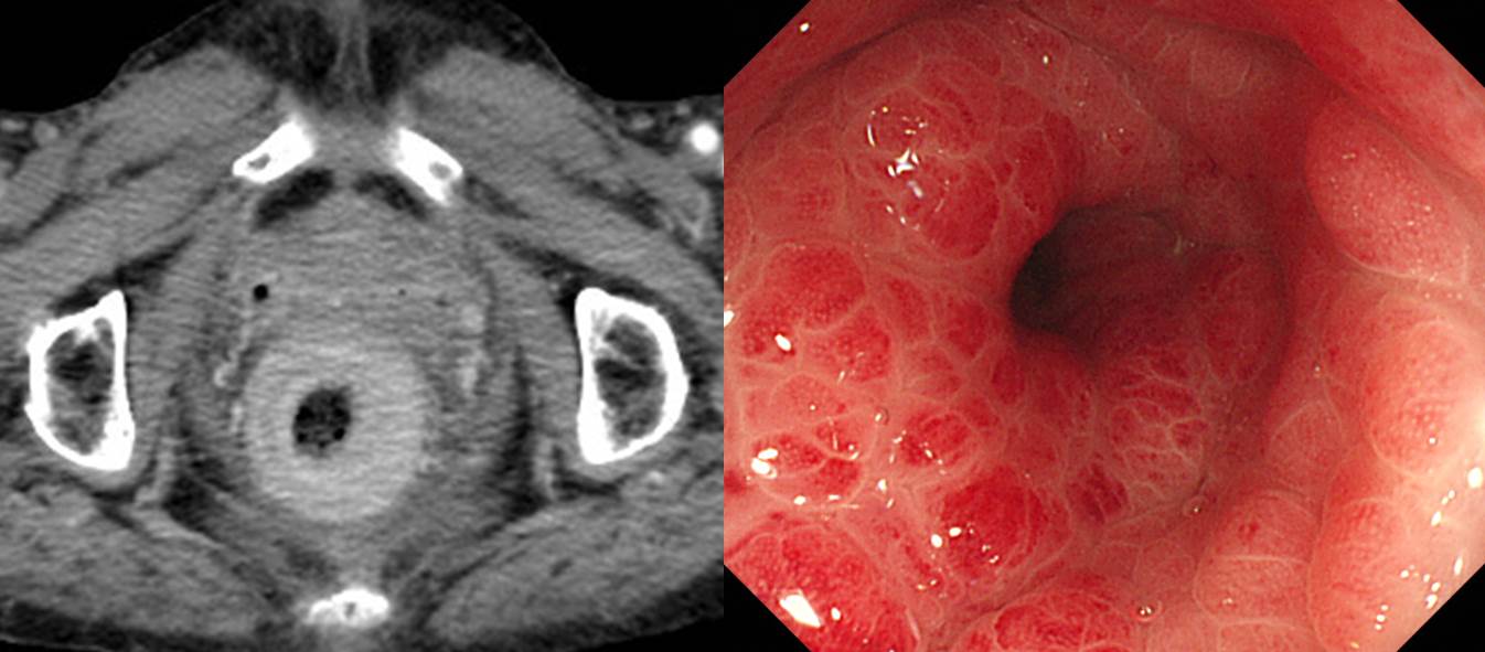
위암 직장 전이 (남/55, 2020)
peritoneal seeding and rectal metastasis (2013. F/65). EGC biopsy; tubular adenocarcinoma, poorly differentiated
위암 total gastrectomy 10년 후 대장 전이 (참고: 위암 수술 후 소견)
조기위암 수술 7년 후 대장 전이와 local recurrence.
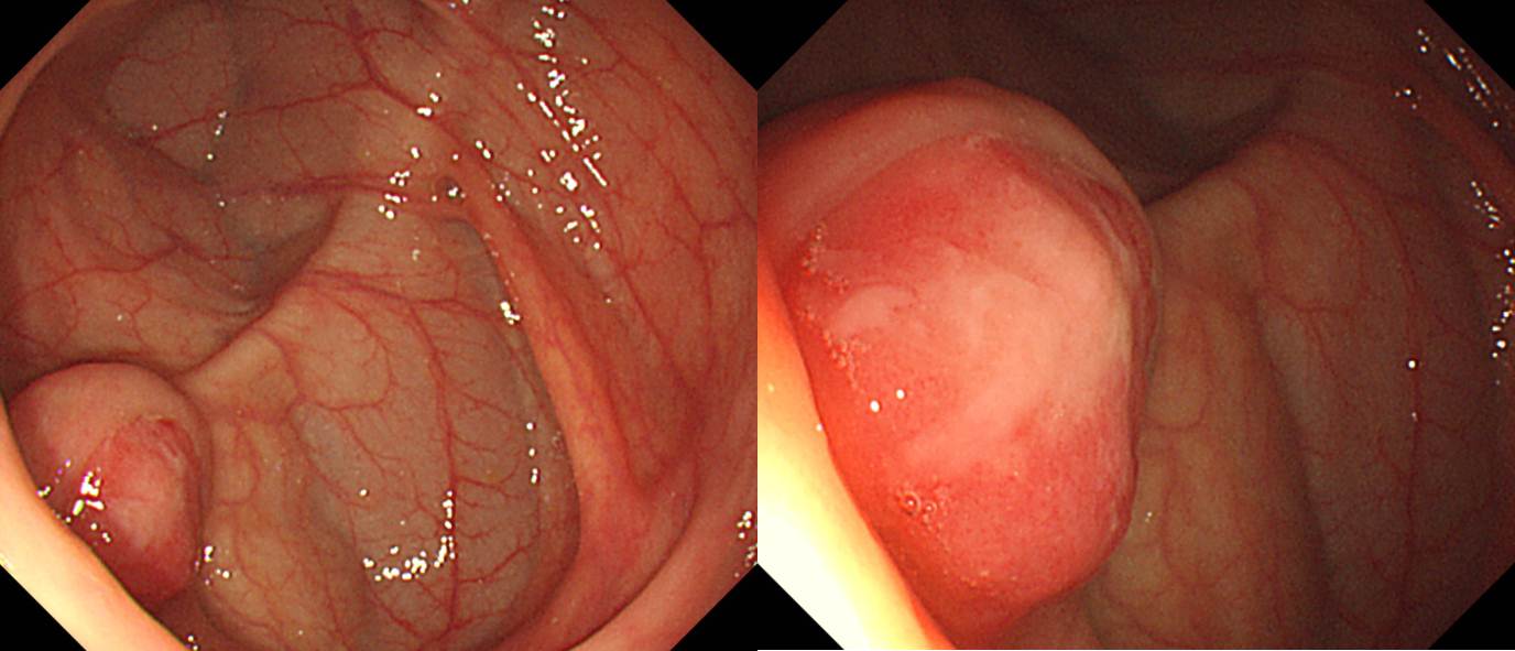
근치적 위암 수술 시행 11년 후 발생한 대장 전이 대한소화기학회지 2020:75:172-175
위암 대장 전이. 두 개의 SMT 양상
다발성 대장암의 다발성 대장 전이 (EndoTODAY 위암 960)
2. 폐암 대장 전이
폐암 대장 전이. Adenoid cystic carcinoma of the lung으로 수술 후 간 전이가 있어 RF ablation 받은 환자의 대장내시경입니다. 수술을 시행하였고 submucosa와 proper muscle layer를 침범하는 metastatic adenoid cystic carcinoma로 확인되었습니다.
폐암 대장 전이. Adrenal gland과 복강내 여러 림프절 전이도 함께 있었습니다.
폐암 대장 전이. 우측 폐의 squamous cell carcinoma, T1aN3M1b, m/pericarium으로 palliative chemotherapy를 하였습니다. 갑자기 발생한 혈변으로 대장내시경을 시행하였고 지혈술을 하였으며 조직검사에서는 poorly differentiated carcinoma가 나와서 수술을 하였습니다. 수술 결과는 아래와 같았습니다.
Transverse colon, transverse colectomy:
Metastatic squamous cell carcinoma:
1) tumor size: 2.7x2.5 cm
2) negative resection margins (safety margin: proximal, 5cm; distal, 12cm)
3. 다양한 암 대장 전이
난소암 대장 전이. Ovary cancer 수술 후 CT 상 rectosigmoid junction 부위가 두꺼워 시행한 대장내시경 조직검사에서 metastatic serous carcinoma가 나왔습니다. (2016년 4월 28일 목요점심내시경집담회)
난소암 직장 전이. 3개의 SMT가 가까이 위치한 모양이었고 조직검사에서는 특이소견이 없었으나 PET에서는 명확한 전이암이 확인되었습니다.
간암 대장 전이.
방광암 대장 전이.
유방암 대장 전이. A 64-year-old woman with a history of stage T2 N1a breast cancer was referred for a screening colonoscopy, which revealed an 8-mm solitary reddish erosion in the sigmoid colon, 28 cm from the anal verge (a). Narrow-band imaging showed dilated small vessels in the depressed area (b). Hematoxylin and eosin staining of the biopsy specimen showed a diffuse infiltrative process in the lamina propria with no dysplastic change in the adjacent colonic epithelium. GATA3 immunohistochemistry confirmed that the infiltrative cells were metastatic breast carcinoma (c). (Am J Gastroenterol 2017;112:677)
[FAQ]
[2017-5-7. 애독자 편지 (제주대 나수영 교수)]
이번 주제(ColonTODAY 대장 전이)와 관련하여 저는 cholangiocarcinoma의 대장 전이를 경험한 적이 있습니다. 사진을 보내드립니다.
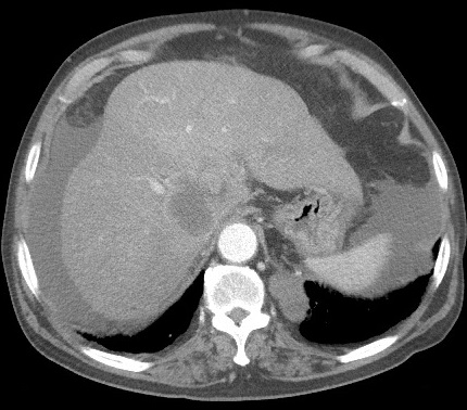
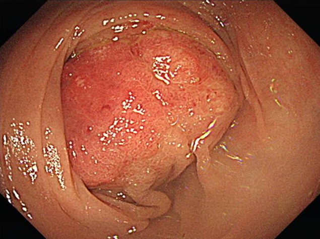
아울러 1990년에 breast cancer로 수술 및 항암 치료 후 2014년에 소장 전이를 포함한 peritoneal metastasis를 보인 환자를 경험한 적이 있습니다. 문헌을 찾아보니 metastatic breast cancer에서 peritoneal metastases는 invasive lobular carcinoma type (68.8%)에서 invasive ductal carcinoma type (1%) 보다 많았다고 합니다 (P = 0.00) (Breast Cancer 2017).
[2025-2-18. 애독자 질문]
직장 및 대장 전이를 보일때, 단순 염증이나, colorectal endometriosis 와 유사한 양상으로 보일 때도 있는데, 조직검사 결과가 확정적이지 않을경우 CT가 도움이 될지요? 혹시 CT 에서도 결과가 확정적이지 않은 경우 추적간격은 어떻게 결정하면 좋을지요?
[2025-2-18. 이준행 답변]
좋은 질문 감사합니다. 어느 정도 강력히 의심되는가에 따라 달라지겠습니다만, 일반적으로 대장 전이가 의심되는데 조직검사에서 확인되지 않으면 CT는 도움될 것 같습니다. 만약 강력히 의심되는데 CT에서 나오지 않으면 PET-CT까지는 해 보고 있습니다. 그래도 명확하지 않으면 보통 2개월 후 재검을 권합니다. 전이된 암은 예후가 매우 나쁠 수 있으므로 6개월이나 1년 후에 검사를 한다면 후회할 일이 생길 수 있습니다.
[References]
1) EsoTODAY - Esophageal diseases
2) SmallTODAY - Small bowel diseases
3) ColonTODAY - Colorectal diseases
4) Dr. Sinn's LiverTODAY - Liver diseases
© 일원내시경교실 바른내시경연구소 이준행. EndoTODAY Endoscopy Learning Center. Lee Jun Haeng.