 EndoTODAY 내시경 교실
Beginner | ESA | Schedule | OPD
Seminars | Atlas | Recent | Links
![]() [Helicobacter 위염] - 終
[Helicobacter 위염] - 終
★ Helicobacter 제균치료 EndoTODAY 표준처방
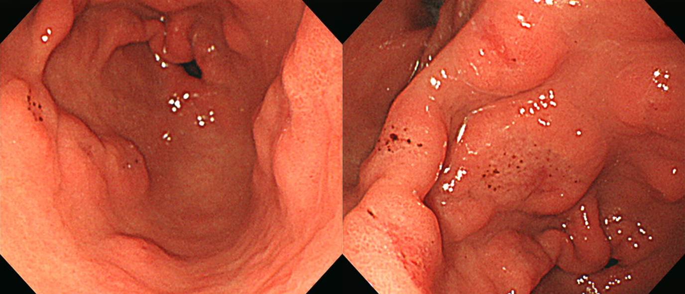
Spotty redness
Spotty redness의 HQ290 near focus 소견
Atrophic and metaplastic gastritis with Helicobacter infection 증례 내시경 동영상
위궤양 증례 내시경 동영상
Hp gastritis
Hp gastritis (active gastritis)
Lymphoid follicle
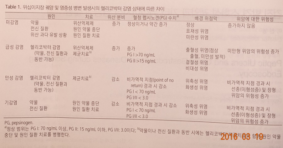
2. Introduction- 헬리코박터 음성 위, 헬리코박터 양성 위
3. 헬리코박터 위염 내시경 소견 - 제균치료 후 호전
4. 헬리코박터 위염에 대한 이선영 교수님과 김광하 교수님의 강의
5. Serum pepsinogen test 와 위축성 위염
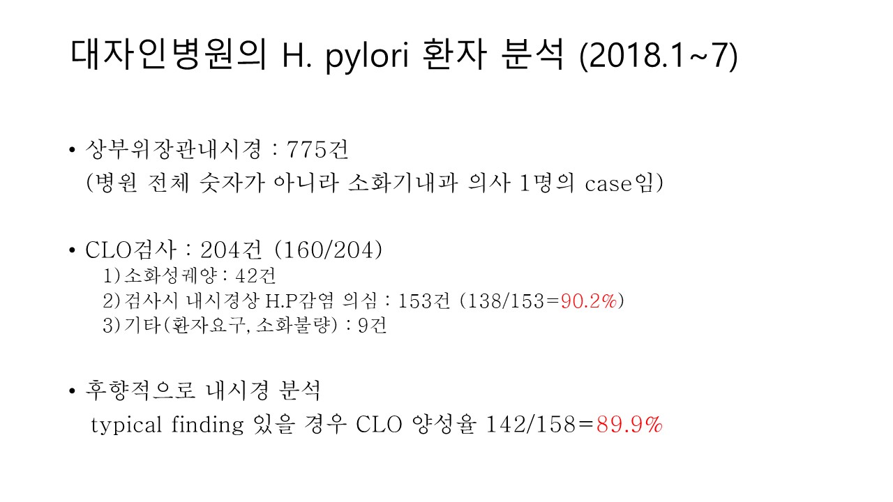
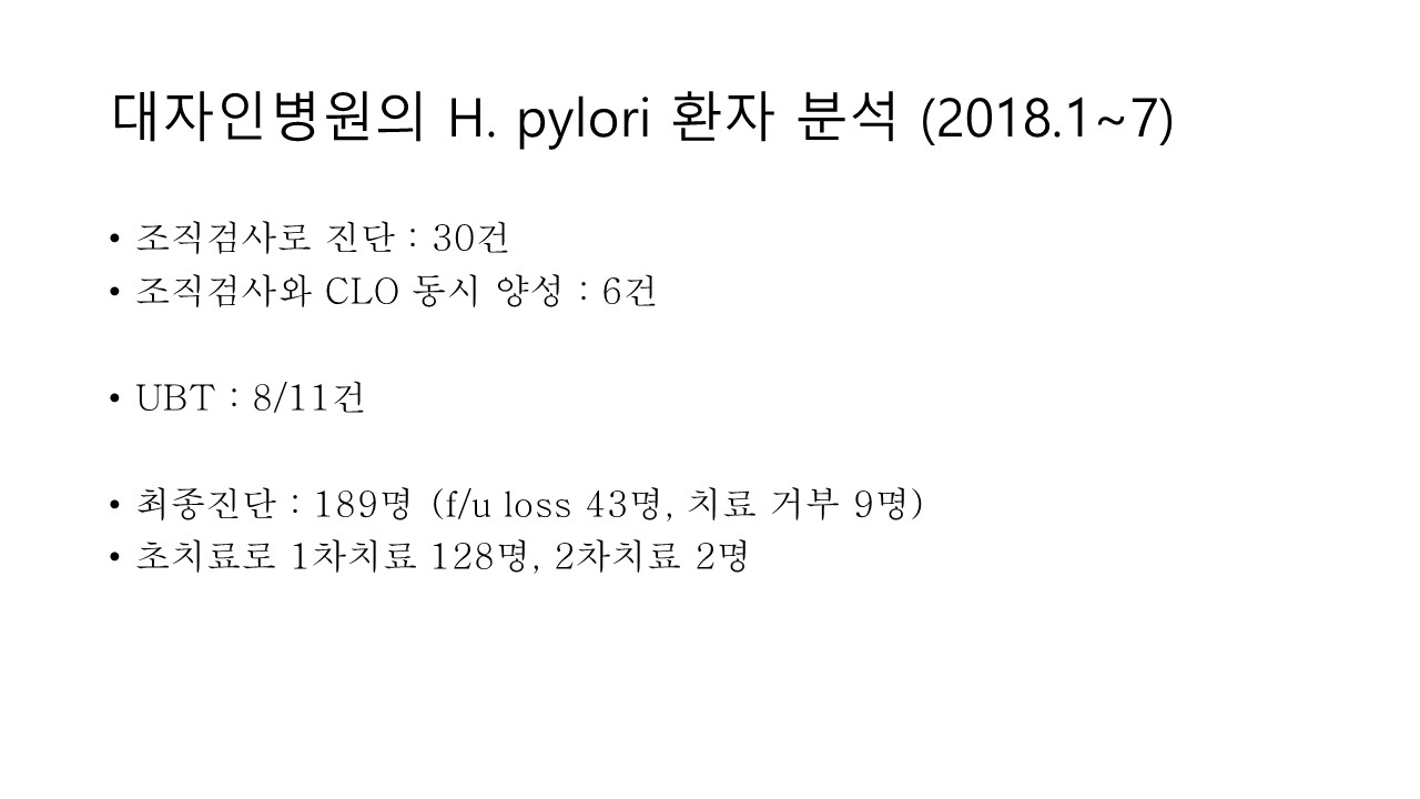
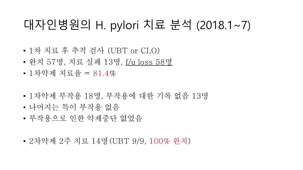
6. 애독자이자 동문이시고 blog '지방의사의 세련된 내과 클리닉' 운영자이신 대자인 병원 내과 노동효 선생님의 '헬리코박터 진단과 치료'
7. Lectures
8. FAQ
9. References

PDF 1.8M. 건국대 이선영 교수님의 위염 quiz 문제와 해설 (2021-9-13)
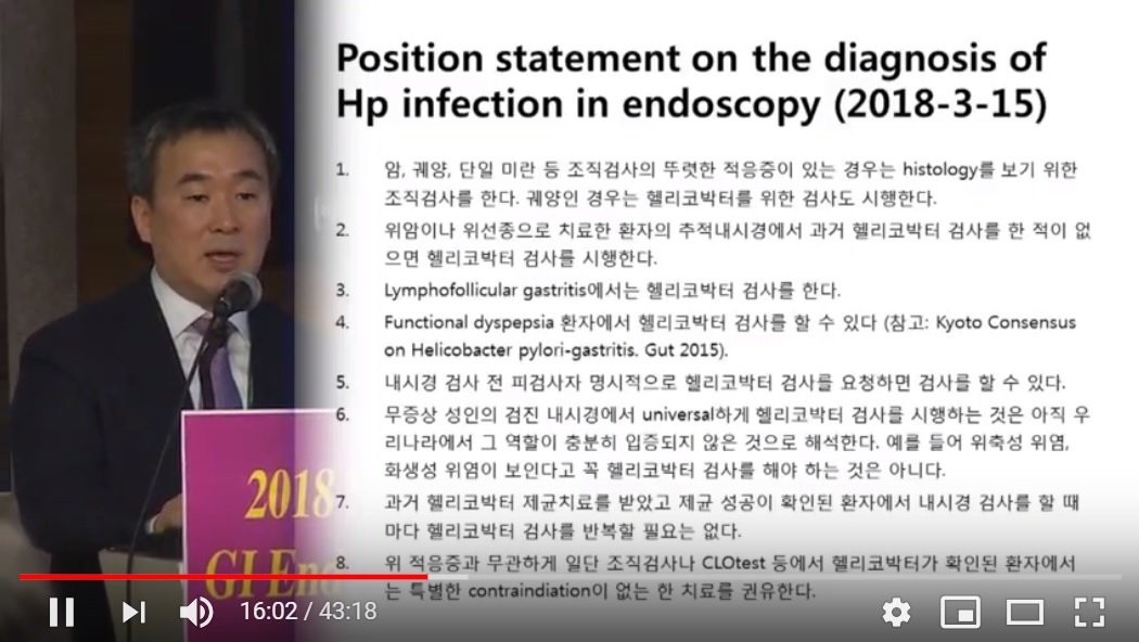
![]() 1. Position statement on Helicobacter gastritis (2018-1-23)
1. Position statement on Helicobacter gastritis (2018-1-23)
위암 발생률 감소를 위하여 헬리코박터 감염이 확인된 환자에서는 제균치료를 시행하는 것이 좋습니다.
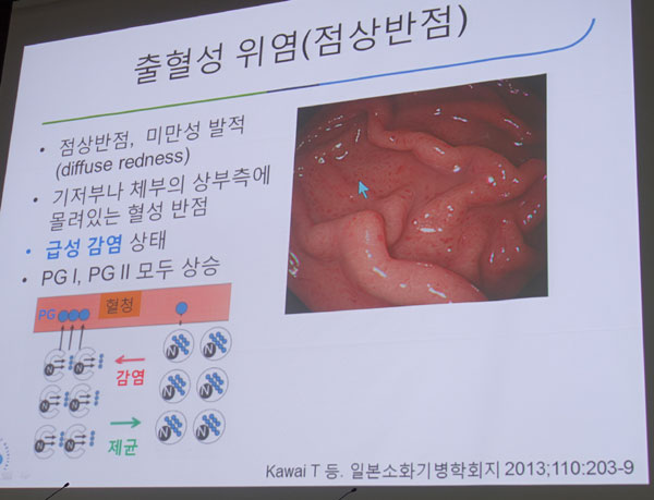
위체상부와 분문부 점막에서 점상 반점, 미만성 발적 (diffuse redness)과 같은 특징적 소견을 보인 환자는 '헬리코박터 위염 (Helicobacter gastritis)'이라는 내시경 진단을 붙이고 몇 개의 조직검사를 할 수 있을 것 같습니다. 만약 헬리코박터 균이 확인되면 제균치료를 시행합니다. 2018년 1월 1일부터 '100/100 급여(약값 전액 환자 부담)'로 치료할 수 있습니다.
2017년까지는 헬리코박터 위염의 제균치료 자체가 불법이었으므로 'chronic superficial gastritis of the proximal stomach (r/o Helicobacter-associated)'라고 애둘러 표현하였지만 (참고) , 이제는 직접적으로 '헬리코박터 위염'으로 표현해도 좋을 것 같습니다. 일본에서 사용하는 용어인 '출혈성 위염'이라는 내시경 진단명은 쓰지 말 것을 권합니다.
![]() 2. Introduction- 헬리코박터 음성 위, 헬리코박터 양성 위
2. Introduction- 헬리코박터 음성 위, 헬리코박터 양성 위
우리나라에서 헬리코박터 위염에 대한 관심이 높아진 것은 2016-7년 무렵입니다. 2015년 EndoTODAY 無答토론 - 만성위염 내시경진단 003를 보면 당시의 우리 나라 내시경계의 현황을 볼 수 있습니다.
헬리코박터가 없는 위는 위축성 변화나 화생성 변화가 없고, 점액이 맑고, RAC이 잘 보입니다.
60대 남성. 헬리코박터 음성. 맑고 깨끗한 위
40대 여성. 헬리코박터 음성. 맑고 깨끗한 위
20대 남성. 검사를 원하여 조직검사와 serology를 했는데 모두 음성
아주 아주 깨끗한 위점막은 어떻게 보일까요.
위 점막의 작은 혈관이 투명하게 비쳐보이면 가장 깨끗한 것이라고 할 수 있겠지요. 그게 바로 RAC (regular arrangement of collecting venules)입니다. RAC에 대한 설명을 ‘상부소화관 내시경진단의 요령과 질환별 내시경상’ 198 쪽에서 요약하여 옮겨보겠습니다. “RAC는 원경에서는 미세한 발적점이 규칙적으로 배열된 모양으로 관찰된다. 근경에서는 불가사리 형태의 미세혈관이다. RAC는 Helicobacter pylori 비감염 위의 체부에서 확인되는 모양이다. 병적 소견이 아니라 위염이 없는 정상 위점막이다.” 같은 책 293 쪽에는 “RAC 음성 증례는 위암을 염두에 두고 내시경 관찰을 해야 한다”라고 언급되어 있습니다. 이는 RAC가 안 보이면 H. pylori가 있는 것이고 그렇다면 위암의 고위험군이므로 위를 자세히 보라는 의미로 해석할 수 있습니다. 믿거나 말거나 수준의 언급이 아닐 수 없습니다. 실제 바쁜 내시경 검사 도중 모든 환자에서 RAC 양성/음성을 따진다는 것은 불가능한 일입니다. 사실 RAC 음성이 위암을 예측하는데 크게 도움이 되는 것도 아닙니다. 주장만 있을 뿐 근거는 거의 없는 소수 일본의사의 私見일 뿐입니다. 요컨데 ‘일본인들이 말하는 RAC라는 것이 있기는 있다. 그리고 가는 혈관으로 보인다’ 정도만 알아두면 충분할 것 같습니다. 헬리코박터와 연관지어 임상적 의의를 크게 부여할 필요는 없을 것입니다. 저는 RAC를 잘 보면 그냥 '깨끗한 위를 봐서 기분이 좋다' 정도 생각하고 넘어가고 있습니다.
2018 APDW (선생님 ?)
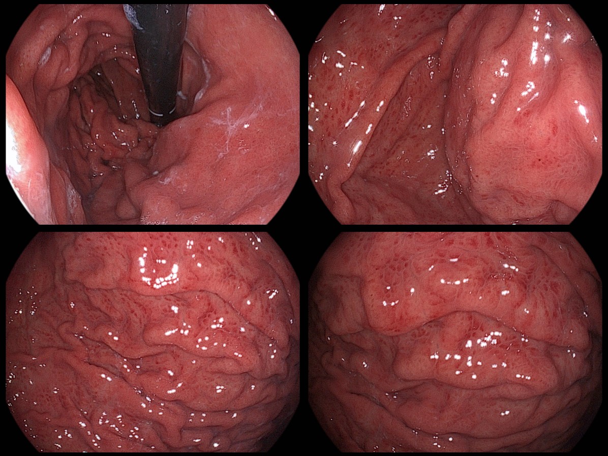
헬리코박터 위염의 가장 특징적인 소견은 위체상부와 분문부 점막의 점상 반점 (spotty redness), 미만성 발적 (diffuse redness), lymphofollicular gastritis, 위점막주름 비후, exudate 등입니다. 만성으로 진행하면 atrophic, metaplastic change를 동반합니다.
우리나라에서는 diffuse redness와 spotty redness는 명확히 구분하지 않고 쓰는 경향이 있습니다. 그러나 2021년 6월 한 심포지엄(2021-6-13. 내시경학회 경인지회 심포지엄)에서 상세히 논의된 바와 이에 대해 이선영 교수님과의 문답을 곰곰히 생각해 보았습니다. 제 결론은 이렇습니다. 일본측 정의가 다소 모호하기는 하지만 우리가 별도의 정의를 만들 필요는 없을 것 같습니다.
- Diffuse redness refers to uniformly reddish mucosa with continous expansion observed in non-atrophic mucosa main in the body.
- Spotty redness is marked by irregular red dots of various shapes and sizes.
아래는 전형적인 증례 사진입니다.
Diffuse redness
일본에서는 '급성 헬리코박터 감염'이라는 용어를 쓰고 있습니다. 그러나 통상의 감염병에서 말하는 수 주 정도라는 의미의 급성이 아니라, 아직 위축성 변화가 오지 않았다는 의미에서의 급성일 뿐입니다. 일본에서는 출혈성 위염이라고 부르고 있으나, 토혈이나 혈변을 일으키는 것은 아니므로 부적절한 용어입니다. 저는 H. pylori-associated gastritis (of the proximal stomach)이라고 부를 것을 제안합니다.
![]() 3. 헬리코박터 위염 내시경 소견
3. 헬리코박터 위염 내시경 소견
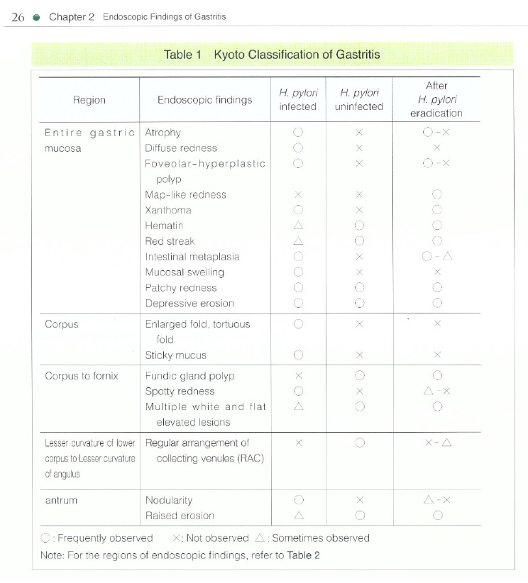
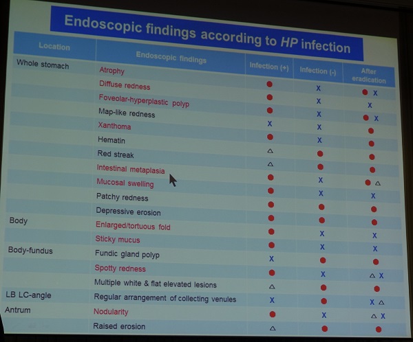
헬리코박터 위염의 내시경 소견에 대해서는 Kyoto classification of gastritis에서 자세히 다루고 있습니다. 위염을 Hp infected, Hp uninfected, After Hp eradication으로 나누어 설명하고 있습니다.
건국대 이선영 교수님의 종설 Endoscopic gastritis, serum pepsinogen assay, and Helicobacter pylori infection Korean J Intern Med 2016에서 관련된 부분을 옮깁니다.
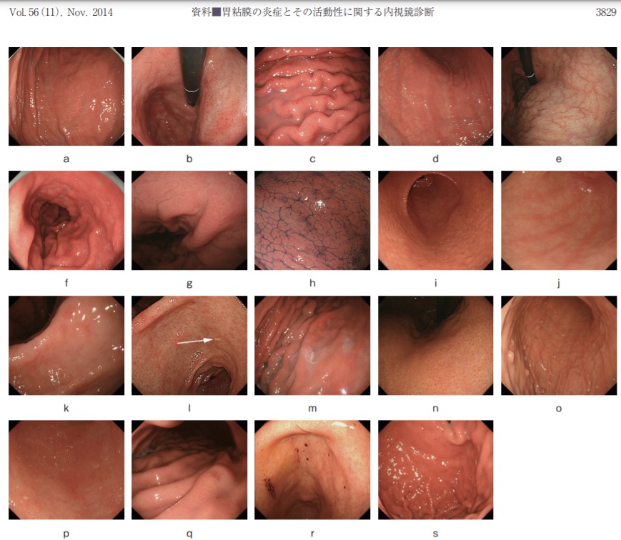
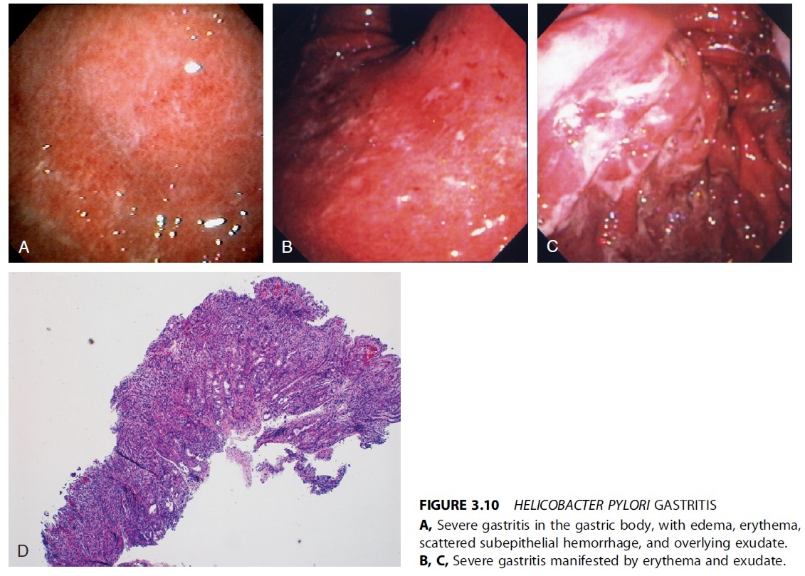
Hemorrhagic spots, nodularity, and thickened gastric folds
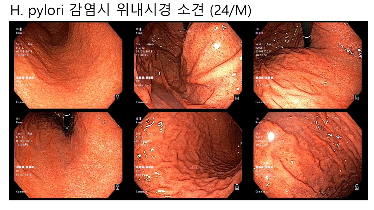
Typical endoscopic findings of acute H. pylori infection include hemorrhagic spots on the fundus and/or high-body, nodular gastritis, and hypertrophic gastric rugae. The endoscopic findings of nodular gastritis are small, round, yellowish-white nodules that represent histological lymphoid follicles. Endoscopic nodular gastritis is more common at the antrum than the corpus in H. pylori infection.
(A) Endoscopic findings in subjects with active Helicobacter pylori infection. (B) Nodular gastritis on the anterior-greater side of the proximal antrum. Multiple small nodules are visible on the antrum, extending up to greater curvature side of the corpus. The nodules consist of submucosal elevated lesions, and thus, there is no color change in nodular gastritis. (C) Follow-up findings of enlarged nodules on the proximal antrum to low-body in the same patient. The previously noted tiny, regular nodules have increased in size. The nodules were irregular and had grown from 12 months prior. (D) Finding of hemorrhagic spots on the fundus in nodular gastritis patient at initial endoscopy (B). Multiple tiny reddish spots, so-called diffuse redness, can be seen on the fundus and greater curvature side of the corpus. (E) Hypertrophic gastric folds. Thickened gastric rugae with whitish, sticky exudates indicate active H. pylori infection. PG, pepsinogen.
어떤 내시경 책에서 헬리코박터 위염을 설명한 부분이 있어서 일부를 옮깁니다.
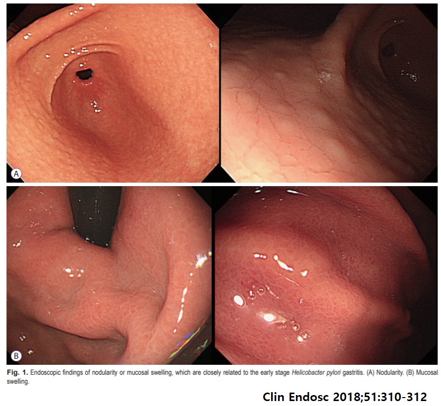
Clinical Endoscopy 2018년 8월호 commentary에서 early stage 헬리코박터 위염의 사진을 소개하고 있었습니다. Nodularity와 mucosal swelling이었습니다. Nodularity는 수구나 수긍할 수 있는 수준이었는데, mocusal swelling은 다소 주관적인 것 같습니다.
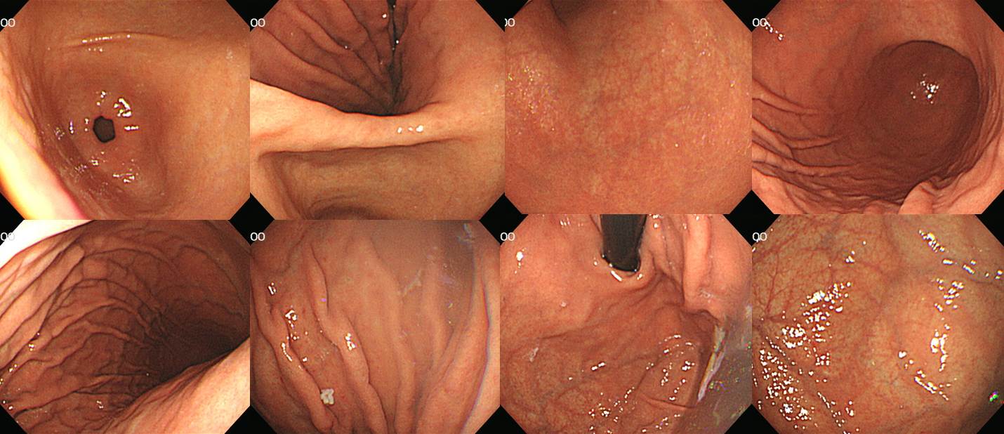
아래는 증례 사진입니다.
Spotty redness
Spotty redness (무답토론 만성위염 3)
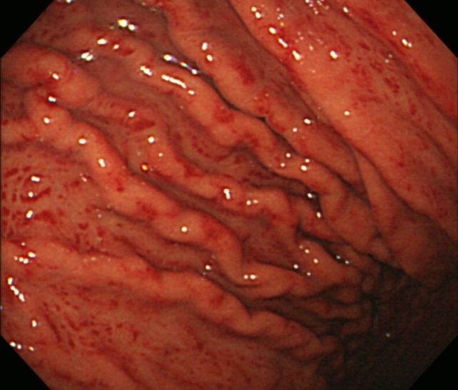
70대 만성 헬리코박터 위염. Atrophy, xanthoma, thick exudate
70대 FD 환자. 소만쪽으로는 넓은 위축성 변화, 위체하부 대만 쪽으로는 linear한 위축성 변화, 위체상부 대만에는 active gastritis (diffuse & spotty redness)
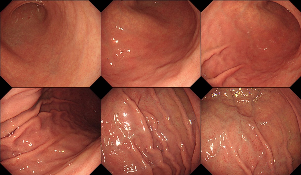
헬리코박터 위염
헬리코박터 위염
헬리코박터 위염
헬리코박터 위염. F/67. 일반적인 감염 경로와 연령, 그리고 일부 atrophic change가 동반되어 있음을 고려할 때 acute라고 말하기는 어려울 것입니다. 그러나 Helicobacter 위염은 틀림 없습니다.
헬리코박터 위염. 비특이적인 상부위장관 증상을 가진 중년 여성입니다. 이런 경우는 환자가 사전에 명시적으로 요청하지 않았더라도 헬리코박터 검사를 하고 일단 제균치료를 시도하는 것이 좋다고 생각합니다. (참고: Kyoto global consensus)
헬리코박터 위염. 혈액 투석 중인 환자입니다. 간질환은 없었습니다.
헬리코박터 위염. 2011년 유방암 수술 환자의 위염
Helicobacter pylori 감염에 의한 비후성 위염
헬리코박터 위염. 한 애독자께서 위벽이 약간 두꺼워 보인 환자에 대하여 아래와 같이 기술하고 조직검사를 하셨다고 합니다. "On the GC of HB, folds were getting thicker and more hyperemic. But this site was similar to that in previous exam." 조직검사는 Hp 위염으로만 나왔습니다. 일단 제균치료를 해 보는 것이 좋겠다고 comment 하였습니다.
헬리코박터 위염. 어떤 전공의 선생님의 내시경 결과지입니다. Helicobacter 제균치료의 제한이 있는 우리나라에서 진단명에 Helicobacter-associated gastritis라고 쓰는 것이 타당한지 논란이 있습니다. 저는 적극적으로 진단하고 치료하는 것이 좋다고 생각합니다. (2017년 8월 당시에는 논란이 있었습니다. 그러나 2018년 제균치료 적응증 확대 이후에는 주저하지 말고 제균치료를 할 것을 권하고 있습니다.)
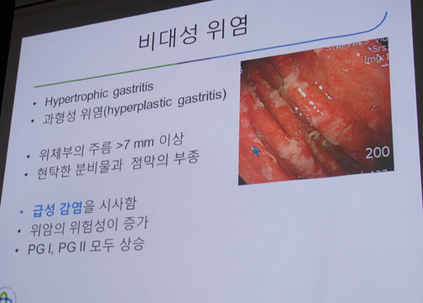
결절성 위염과 위주름의 아주 경미한 비후. 헬리코박터 위염에서 위주름이 약간 두껍다는 것은 대부분 이 정도의 경미한 소견입니다. 명백한 비후성 위염은 흔하지 않습니다.
비후성 위염 형태의 헬리코박터 위염
Helicobacter-associated hyperplastic polyp
전정부 위암 + Helicobacter gastritis
위전정부 점막하종양이 두 개 있으면서 lymphofollicular gastritis 소견이 보였습니다.
위전정부 고도선종 + 헬리코박터 위염
78세 여성의 전형적인 헬리코박터 위염입니다. 약간의 hiatal hernia와 columnar-lined esophagus 소견이 있었습니다. 산역류 증상은 없었습니다.
SMT 의심으로 오신 30대 여성. 위전정부에는 경미한 nodular gastritis, 위저부에는 diffuse redness, 위체부 주름이 다소 두껍고 thick exudate가 관찰되는 전형적인 헬리코박터 위염이었고, SMT라고 오신 곳은 single verruca처럼 보였음
위축성 변화 + 탁한 위액
3차 제균치료 실패. Thick한 exudate.
Fundus의 퇴색된 그물망 무늬로 보이기도 합니다.
F/42 (2020)
위 환자의 조직검사. 내시경 소견에서 lymphofollicular gastritis가 뚜렷하지 않더라도 조직검사에서 lymphoid follicle은 뚜렷할 수 있습니다.
남자 50세 (조직검사)
[Thickened folds]
MALT 림프종을 배제할 수 없다는 소견으로 의뢰되었는데 재검에서는 H. pylori gastritis with lymphoid follicle로 나옴.
Thickened fold로 보만 4형 진행성위암 의심으로 의뢰되었으나 H. pylori 관련 주름 비후로 판단되었음.
[Salt and pepper]
이선영 교수님의 명저 '위염의 내시경소견' 55쪽에 이런 언급이 있습니다. "H. pylori 감염이 지속되면 작은 결절들은 납작해지면서 희끗희끗한 소금 후추가루 양상 (salt-and-pepper)으로 변해간다. 흰 부분들이 뭉치면 점막하 혈관상이 투영되는 위축성 위염으로 보이기 시작한다." Lymphofollicular gastritis (= 결절성 위염) 이 오래되면 위축성 위염으로 변해가는데 그 과정에 salt and pepper 점막이 보인다는 것입니다. 아래와 같이 보입니다.
위체하부 소만의 salt and pepper 양상과 위전정부의 lymphofollicular gastritis
위체하부 소만의 salt and pepper 양상과 위전정부의 위축성 변화
수 년 전 한 일본 연구(JGH Open 2018;2:80-86)에서 corpus white spots라고 언급한 부위도 이러한 소견을 말하는 것으로 생각됩니다.
Endoscopic appearance and pathological features of nodular gastritis. (a) and (b) are typical endoscopic images of nodular gastritis (NG). (a) An unusual miliary pattern resembling “gooseflesh” in the antrum. (b) Antral nodularity is highlighted with chromoendoscopy using indigo?carmine dye spraying. (c) A pathological finding of biopsy specimens from NG. Pathological findings from biopsy specimens are characterized by superficially located prominent lymphoid follicles with a germinal center (hematoxylin and eosin staining). (d) and (e) White spot aggregates in gastric corpus (corpus white spots: CWS) in a patient with NG. A striking result of this study was the link between CWS and risk of cancer development in patients with NG. The histological findings of CWS, examined via surgical specimens, included hyperplasia of lymphoid follicles with intense mucosal inflammation, similar to NG. Patients with NG and CWS have highly active inflammation of the corpus, which is implicated in the development of diffuse?type cancer. Consequently, the presence of CWS, observed during upper endoscopy for patients with NG, should be dutifully noted. Patients who display CWS should undergo urgent H. pylori eradication therapy. (JGH Open 2018;2:80-86)
한 연구에 따르면 헬리코박터 제균치료를 하지 않으면 "작은 과립형의 결절성 위염은 소금-후추가루 양상을 거쳐 위축성 위염으로 진행하는 경향을 보였으며, 큰 결절형은 불규칙한 점막의 융기로 구성된 화생성 위염으로 진행하는 경향을" 보입니다. (Korean J Gastroenterol 2019)
Lymphofollicular gastritis (= 결절성 위염)과 salt and pepper 양상 위점막이 보이는 경우 헬리코박터를 검사하고 치료하시기 바랍니다.
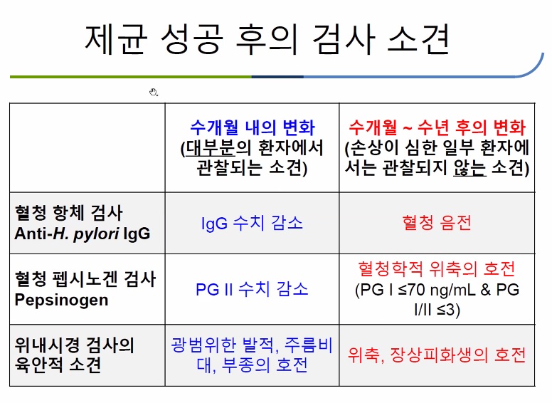
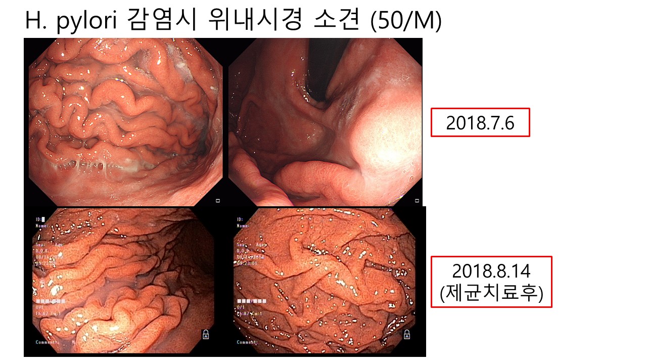
[제균치료 후 호전]
헬리코박터 제균치료 후 atrophy나 metaplasia는 천천히 호전되지만 spotty redness와 같은 소견은 비교적 금방 호전됩니다.
Spotty redness와 edematous fold가 제균치료 1년 후 현저히 호전됨
Thickened fold로 보만 4형 진행성 위암 의심으로 의뢰되었으나 헬리코박터 제균치료 후 현저히 호전
좌측은 제균치료 전 Olympus 내시경 사진, 우측은 제균 1년 후 Pentax 내시경 사진이지만 제균치료 후 현저히 호전되었음을 알 수 있습니다.
제균치료 후 현저한 호전
제균치료 2개월 후 현저한 호전
제균치료 후 현저한 호전
제균치료 5년 후 위점막. 헬리코박터 위축성 위염에서 atrophic border의 바깥쪽은 염증이 심해 보이기 마련입니다. 제균치료를 하면 염증은 줄어드는데 위축은 호전되지 않는 경우가 있습니다. 그러면 atrophic border 바깥의 위점막이 너무나 깨끗하게 보일 수 있습니다.
Tokyo classification of gastritis (2017) 55쪽. 제균치료 후 초기에는 small granular elevation은 flattened 되지만 white spot은 계속 보일 수 있습니다. 오래되면 위축성 위염이 남는다고 합니다.
제균치료 후 보이는 소견 중 유명한 것이 map-like redness입니다. Atrophy가 있던 사람에서 치료 후 염증만 있던 부위와 atophy가 있던 부위의 호전 양상이 달라서 발생하는 것입니다. Map-like redness가 있는 경우 암 발생이 많았다는 보고가 있습니다 (Dig Endosc 2016). Patchy redness와 whitish elevated lesions도 성공적인 제균 후 보일 수 있는 소견입니다.
[위암 + 헬리코박터 위염]
위체부 pale한 조기위암(signet ring cell carcinoma) 환자입니다. 위체상부와 fundus에 전형적인 Helicobacter 위염이 있었습니다.
30대 여성. Signet ring cell carcinoma. Fundus 사진을 보면서 제 가슴이 찢어졌습니다. 빨리 헬리코박터 제균치료 적응증을 확대해야 할 것 같습니다. 안타까운 사연이 너무 많습니다.
유방암 수술 5년 후 위암이 진단되었습니다. Fundus의 헬리코박터 위염이 현저하였습니다.
매우 작은 signet ring cell carcinoma + 헬리코박터 위염
전정부 전벽의 lymphofollicular gastritis가 뚜렷했습니다. Signet ring cell carcinoma
젊은 여성 위암 + 헬리코박터 위염
위암 + 헬리코박터 위염
위암으로 의뢰된 40세 여성입니다. 의뢰 받을 당시 위체부 미분화조직형 위암으로만 의뢰되었습니다. 그런데.... 제가 보니 Helicobacter 위염이 너무나 뚜렸습니다. 위암 예방을 위한 헬리코박터 제균치료가 꼭 필요다고 생각합니다.
Stomach, radical subtotal gastrectomy:
. Early gastric carcinoma
1. Location : middle third, Center at body and posterior wall
2. Gross type : EGC type IIc
3. Histologic type : tubular adenocarcinoma, poorly (solid) differentiated with mucinous component (30%)
4. Histologic type by Lauren : diffuse
5. Size : 5.4x5 cm
6. Depth of invasion : invades submucosa (sm3) (pT1b)
7. Resection margin: free from carcinoma, safety margin: proximal 2.5 cm, distal 7 cm
8. Lymph node metastasis : no metastasis in 51 regional lymph nodes (pN0)
9. Lymphatic invasion : not identified
10. Venous invasion : not identified
11. Perineural invasion : not identified
12. Peritoneal cytology : negative
13. AJCC stage by 7th edition: pT1b N0
작은 signet ring cell carcinoma + 헬리코박터 위염
MALToma + 헬리코박터 위염
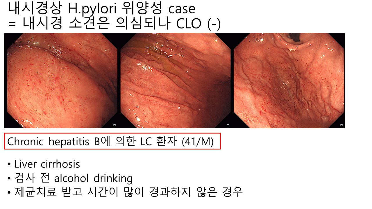
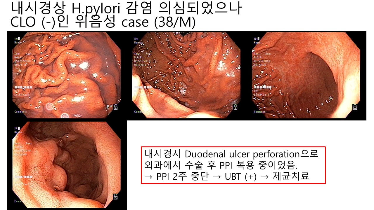
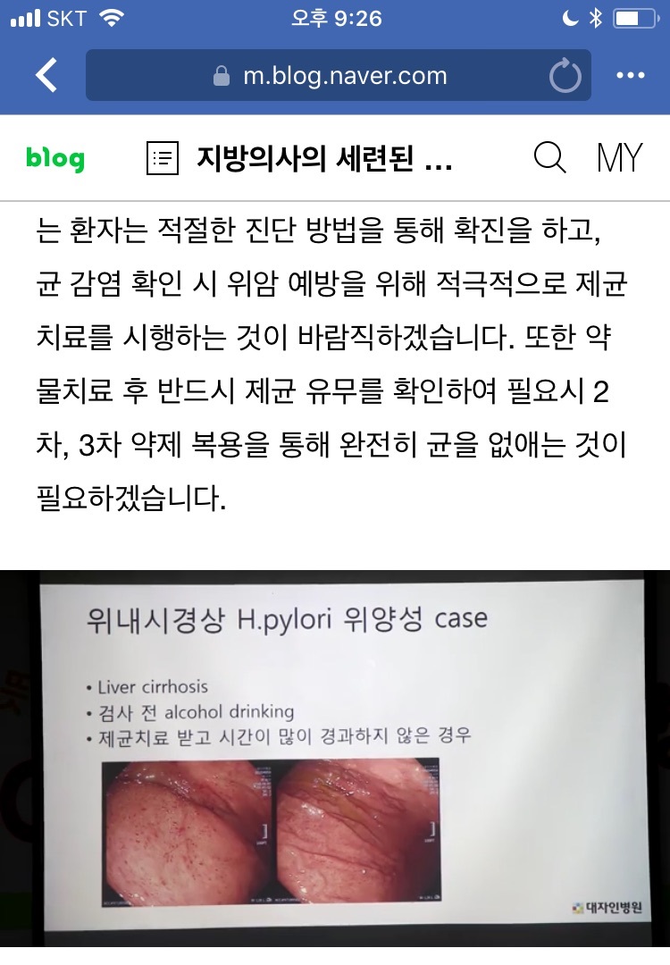
헬리코박터 위염과 비슷한 소견이지만 실제로 헬리코박터 감염이 없는 경우가 있습니다. Facebook에서 대자인 병원의 노동효 교수님의 강의 동영상을 보면서 좋은 증례가 있어 소개합니다.
헬리코박터 위염에 대하여 고민하면서 진료하고 있습니다. 내시경으로 헬리코박터 감염 여부를 대충 알 수 있다지만...... 실제 임상에서는 애매한 경우가 매우 많습니다. 아래 환자의 경우는 어떻게 생각하십니까?
양성이었습니다. 예, 그렇습니다. 봐도 봐도 잘 모르겠습니다. 뚜렷한 경우도 있지만 애매한 경우가 너무 많은 것이 사실입니다.
아래 사진도 헬리코박터 위염입니다. 멀리서 대강 보면 잘 알 수 없으나 가까이서 high body와 fundus를 보면 헬리코박터 위염이라는 뚜렷한 증거가 있습니다.
[2018-12-7. 이준행 혼잣말]
"내시경 결과지에 Helicobacter 위염이라 씌여 있는데 헬리코박터 검사는 되어 있지 않은 경우를 보았으니 의견을 정리해 달라"는 요청을 받았습니다.
(1) 2018년 1월부터 헬리코박터에 대한 검사가 가능해졌다지만, 특별한 경우(위궤양, 십이지장 궤양 등)를 제외하면 '선별 급여'입니다. 환자가 검사비 대부분을 자비로 내야 합니다. 검사비를 자비로 내야 하는데 사전 동의는 필요하겠지요. 그다지 비싸지 않다면 사전 동의 없이 선별 급여 검사를 해도 좋다고 생각하는 분도 계시지만 원칙은 아니겠습니다.
(2) 우리나라에서는 내시경으로 헬리코박터 감염을 진단하기 위한 시도를 해 본 역사가 일천합니다. 아주 최근에야 학술 모임에서 헬리코박터 위염의 내시경 진단을 논하고 있을 뿐, 몇 년 전까지만 해도 전혀 들을 수 없었습니다. Diffuse redness라는 용어를 잘 모르는 분도 많습니다. 결과지에 "헬리코박터 위염"이라고 씌여 있으면 어떻게 해야 좋을지 정해진 바도 없습니다.
(3) 내시경 육안 소견에 따라 결과지에 "헬리코박터 위염"이라고 쓰면, 이를 근거로 헬리코박터 제균치료(이 또한 선별급여입니다)를 할 수 있는지도 의문입니다.
따라서 2018년 12월 현재에는 내시경 소견만을 근거로 내시경 결과지 impression 란에 '헬리코박터 위염'이라 쓰는 것은 다소 성급한 것 같습니다. 꼭 쓰고 싶으면 chronic gastritis (r/o Helicobacter-associated) 정도의 애매한 표현이 낫겠습니다.
"결과지에 'chronic gastritis (r/o Helicobacter-associated)'라고 쓰면 반드시 헬리코박터 검사를 해야 하는 것 아닌가?" 질문하는 분이 계실 것 같습니다. 맘대로 하십시오. 필요하면 언제든지 헬리코박터 검사가 가능한 제도면 좋겠지만, 현행 규정은 '선별 급여'이기 때문에 일관된 방침을 정할 수 없습니다. 검사가 끝난 후 환자가 "왜 비보험 검사를 허락없이 했는가?"라고 물었을 때 잘 설명할 자신이 있으면 (= 경우에 따라 돈을 물어낼 생각이 있으면, 아주 가끔 멱살도^^) 사전 동의 없이 검사를 해도 좋습니다. 그럴 자신이 없으면 검사하지 않아도 좋습니다. 아직까지는 선별급여는 비보험으로 이해하는 환자들이 많기 때문입니다. "90%를 내가 돈을 내야 한다면 그게 비보험이지 왜 보험이냐?"라는 환자의 질문은 타당한 문제제기입니다.
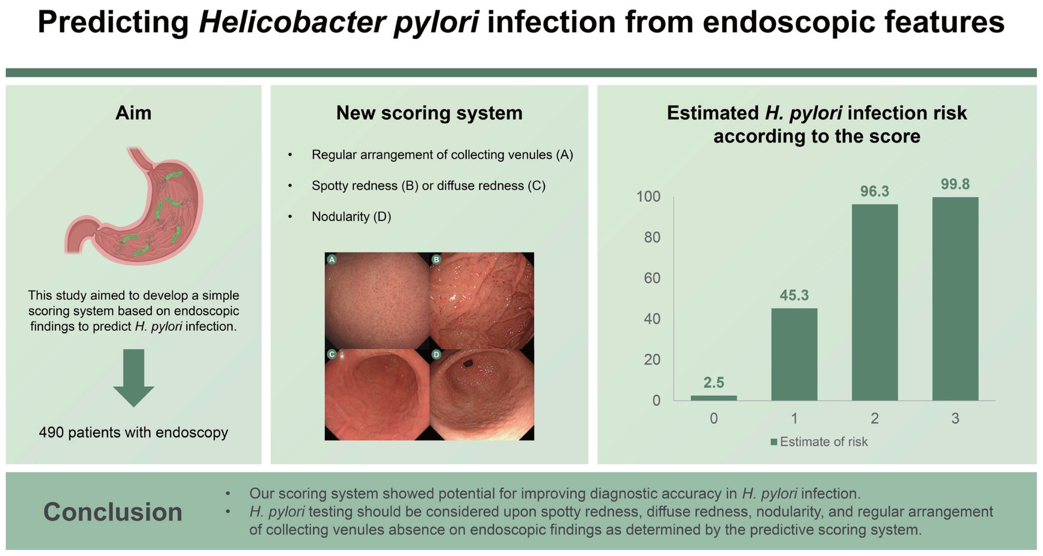
아산병원에서는 simple scoring system을 만들었습니다.
![]() 4. 헬리코박터 위염에 대한 이선영 교수님과 김광하 교수님의 강의
4. 헬리코박터 위염에 대한 이선영 교수님과 김광하 교수님의 강의
1) 2016년 3월 20일 내시경학회 세미나에서 건국대 이선영 교수님께서 헬리코박터 감염의 단계에 따른 위점막 변화를 schematic하게 설명하셨습니다.
2) 2016년 12월 3일 헬리코박터 심포지엄에서 김광하 교수님께서 일본에서 이해하고 있는 Helicobacter gastritis의 내시경 소견을 표로 보여주셨습니다.
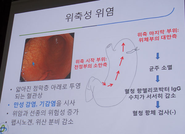
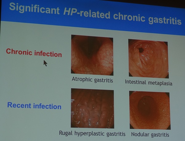
Helicobacter 감염 여부에 따라 내시경 소견에 차이가 있습니다. Chronic infection은 위축성 위염과 화생성 위염, recent infection은 rugal hyperplastic gastritis, nodular gastritis의 소견을 보입니다. (참고: EndoTODAY 비후성 위염)
3) Helicobacter 위염: 내시경으로 진단할 수 있을까? 이선영 (2018년 3월 25일 내시경학회세미나)
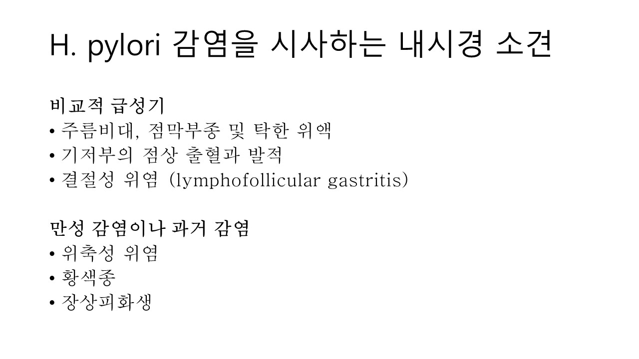
이선영 교수님은 비교적 급성기의 활동성 헬리코박터 위염에 대하여 자세히 설명해 주셨습니다. (1) 주름비대, 점막부종 및 탁한 위액, (2) 기저부의 점상 출혈과 발적, (3) 결절성 위염 (lymphofollicular gastritis, 작은 과립형은 위축성 위염으로 진행하는 경향이 있고 큰 결절형은 화생성위염으로 진행하는 경향이 있습니다)입니다. 헬리코박터 감염이 보다 오래된 경우는 (4) 위축성 위염, (5) 황색종, (6) 장상피화생 (지도상 발적, 함몰형 발적은 장상피화생의 소견임) 등이 관찰됩니다.
(2016)
제가 '비교적 급성기의 활동성 헬리코박터 위염'에 대하여 관심을 가지고 있는 이유는 EndoTODAY 헬리코박터 조직검사에 대한 position statement, version 2018-3-15)의 조직검사 적응증에 '결절성 위염'은 포함시켰지만, '(1) 주름비대, 점막부종 및 탁한 위액'과 '(2) 기저부의 점상 출혈과 발적'을 포함시키지 않았기 때문입니다. 이선영 교수님께서는 (1)과 (2)도 포함시키는 것이 좋겠다고 말씀주셨는데 저도 마음 속으로 동의하고 있습니다.
그러나 우리나라 내시경 의사들이 아직까지는 '비교적 급성기의 활동성 헬리코박터 위염'에 익숙하지 않고, 특히 (1) 주름비대, 점막부종 및 탁한 위액, (2) 기저부의 점상 출혈과 발적을 헬리코박터 감염과 연결시켜온 전통이 없습니다. 우리나라 내시경 의사들이 이에 대한 개념과 경험을 갖게 된다면 저도 position statement를 변경할 예정입니다. 매우 중요한 topic에 대하여 compact하고 충실한 강의를 해 주신 이선영 교수님께 감사드립니다.
![]() 5. Serum pepsinogen test 와 위축성 위염
5. Serum pepsinogen test 와 위축성 위염
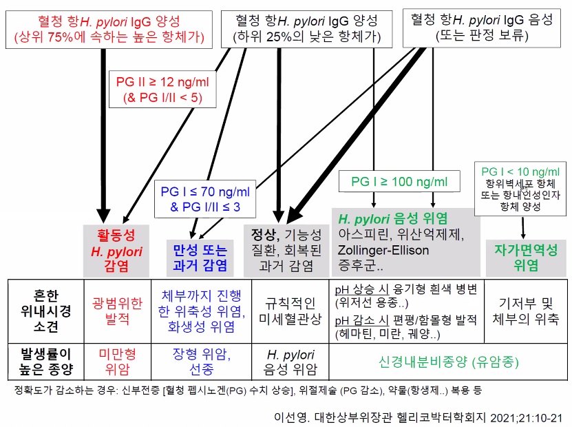
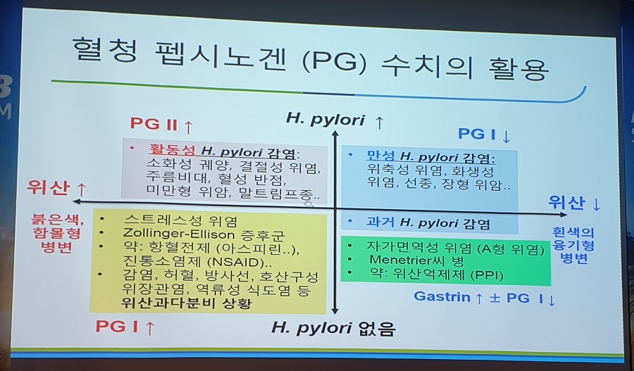
[H. pylori 감염과 serum pepsinogen test]
이선영 교수님의 논문 (Endoscopic gastritis, serum pepsinogen assay, and Helicobacter pylori infection. Korean J Intern Med 2016)에서 옮깁니다.
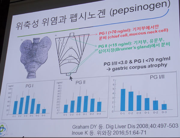
정상 위입니다. PG I은 주로 fundus에서 분비되고 PG II는 전 stomach에서 분비됩니다.
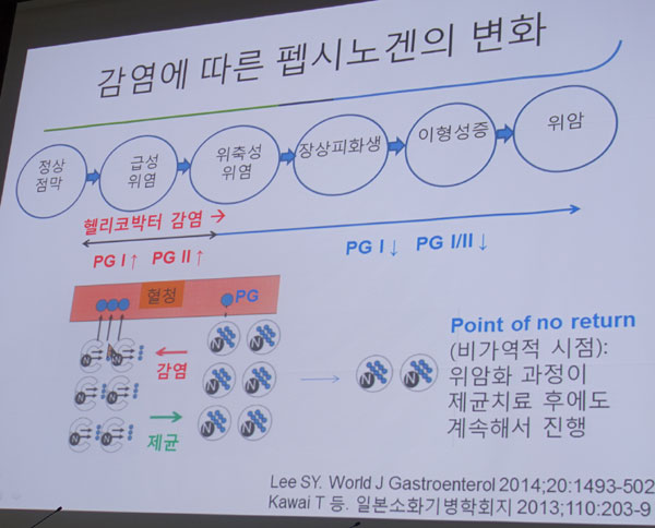
Helicobacter pylori 감염이 오래 되지 않은 상태입니다. 아직 위축성 변화가 오지 않았고 염증성 변화만 많은 상태입니다. PG 분비는 오히려 높아진 상태입니다.
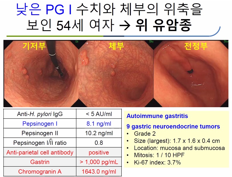
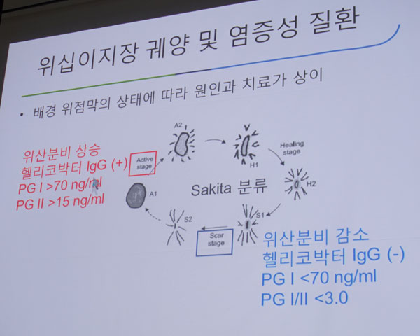
Helicobacter pylori 감염이 만성화되면 위축성 변화가 현저해집니다. PG I과 PG II가 모두 줄어드는데, PG I이 상대적으로 더 많이 줄어들어 PG I/II ratio도 낮아집니다.
Gastric corpus atrophy is defined as a serum PG I/II ratio of < 3.0 and a serum PG I level of < 70 ng/mL. Although endoscopic, histological, and serological atrophic gastritis are well correlated, the presence of gastric corpus atrophy is not always consistent with CAG found by endoscopy. When histological atrophic gastritis progresses, serum PG I levels and the PG I/II ratio are decreased. Similarly, when the extent of atrophy is increased, the serum PG I/II ratio decreases. Moreover, the serum PG I/II ratio is significantly decreased in subjects with severe or moderate CAG than in those with mild CAG.
[2021-4-17. 순천만내시경세미나 이선영 교수님 강의- 위염에 대한 혈청검사]
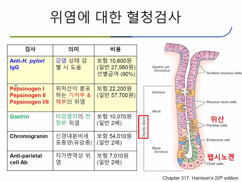
Anti-Helicobacter 항체가 양성이라도 수치가 낮으면 false positive인 경우가 많습니다.
anti-Helicobacter 항체 수치가 높았던 사람이 제균치료 후 항체 수치가 상당히 낮아지면 비록 양성이라고 하더라도 헬리코박터 제균치료 성공임을 의미합니다.
PG-II가 헬리코박터에 도움이 되고 PG-I는 위축성 위염에 도움이 됩니다.
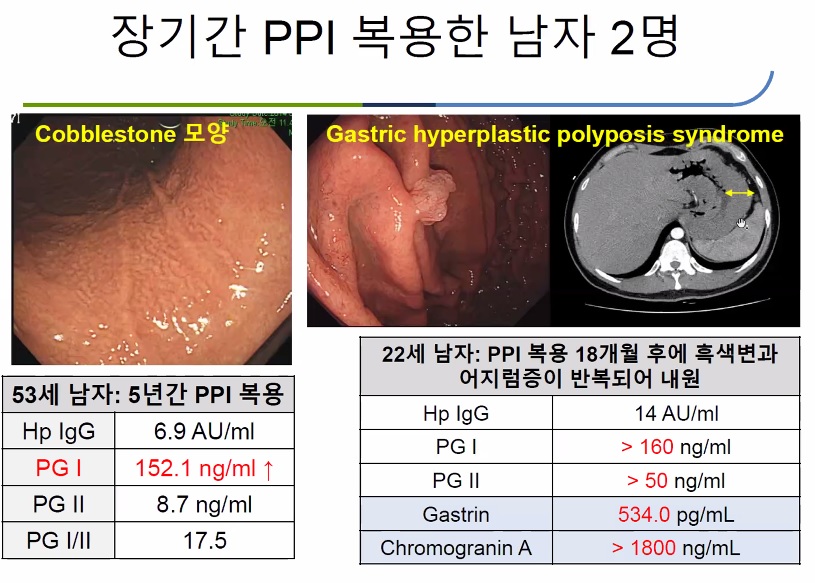
PPI 사용시 여자에서는 납작한 fundic gland polyp이 많이 발생하고, 남자에서는 cobblestone appearance를 보일 수 있습니다.
[2013-7-8. 애독자 질문]
위축성 위염 정도만 있어서는 CLOtest 양성이어도 제균치료를 권장하시는지요?
[2013-7-8. 이준행 답변]
의료진마다 생각이 다른 분야입니다. 저는 균이 나오면 꼭 치료해야 하는 상황에서만 CLOtest를 시행합니다. 위궤양이나 십이지장궤양, MALT 림프종 등입니다. 위축성 위염에서는 CLOtest를 시행하지 않습니다. 이러지도 저러지도 못하는 상황이 되기 쉽기 때문입니다. 우연히 위축성 위염 환자에서 헬리코박터 양성이 나와도 치료를 먼저 권하지는 않습니다.
그리고 위축성 위염에서 헬리코박터를 검사하는 것 자체가 현행 규정 밖입니다. 이유가 명확한 경우에는 규정밖이라도 CLOtest를 해볼 수 있다고 생각합니다. 그러나 저는 가급적 현행 규정을 지킬 것을 권합니다. 많은 연구와 consensus meeting 등을 통해 규정을 바꾸기 위해 노력하는 것이 옳습니다. 의사 개개인이 나름의 생각으로 너무 쉽게 규정을 위반하는 것은 바람직하지 않다고 봅니다. 이로 인한 환자들의 혼선도 큽니다. 병원마다 의사마다 다르면 환자는 헷갈리기 마련입니다. 보통 좋지 않은 방향으로 결론이 납니다.
[2018-6-17. 이준행 수정 답변]
2018년 1월 1일부터 헬리코박터 제균치료가 적응증과 무관하게 합법화 되었습니다. 2018년부터는 헬리코박터 감염이 확인된 위축성 위염에서는 제균치료를 권하고 있습니다.
![]() 6. 애독자이자 동문이시고 blog '지방의사의 세련된 내과 클리닉' 운영자이신 대자인 병원 내과 노동효 선생님의 '헬리코박터 진단과 치료'
6. 애독자이자 동문이시고 blog '지방의사의 세련된 내과 클리닉' 운영자이신 대자인 병원 내과 노동효 선생님의 '헬리코박터 진단과 치료'
교수님, 전화를 받고 정말 감사했습니다.
병원을 떠난지 몇년 지난 제자를 기억해 주시고, 부족한 저의 발표까지 봐 주셔서 감사합니다. 교수님의 EndoTODAY는 2weekly 또는 monthly로 정독하고 있습니다. 비록 제가 대학병원을 떠났지만, 교수님께서 항상 말씀하신대로 바른 내시경을 하기 위해서 노력하고 있습니다. 언제 어디서든 삼성서울병원 소화기내과에 누가 되지 않는 소화기의사가 되겠습니다.
핵심 슬라이드만 그림 파일로 만들어보았습니다. 감사합니다.
1) 2019-10-13. 이선영 교수님 강의
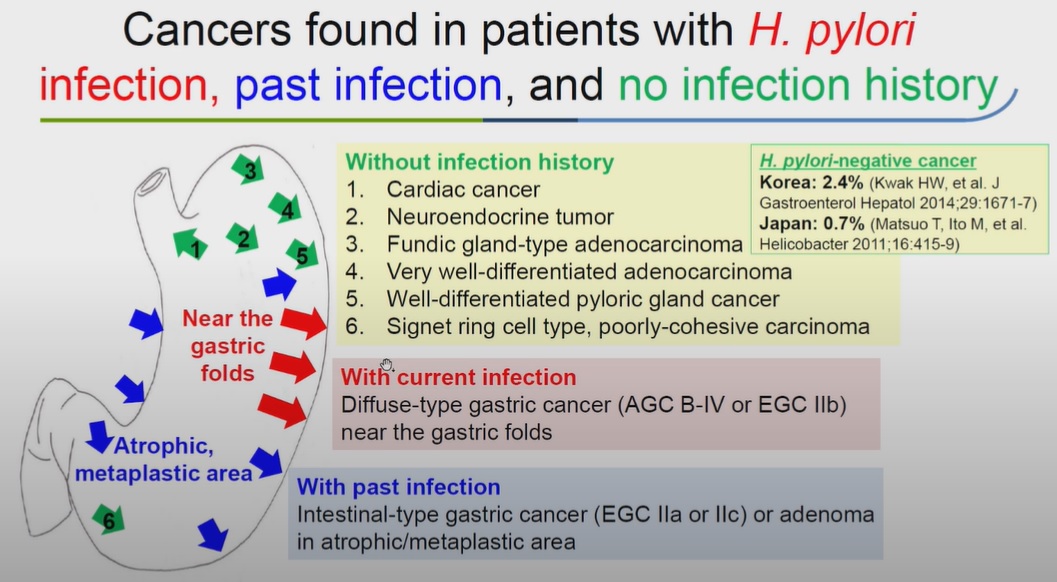
고령자의 위암은 줄고 있으나 젊은 환자의 위암은 늘고 있습니다. 1차 예방을 위한 노력이 필요함을 보여주는 자료입니다.
True Hp 음성 위암의 절반이 근위부 위암입니다.
조직검사의 sensitivity는 충분하지 않으므로 대변 검사를 추가하는 것을 추천하고 싶습니다. 그러나 우리나라에서는 워낙 처방례가 적어서 제조사가 생산 중단을 고려하고 있는 정도라고 합니다.
Yes/No뿐만 아니고 수치로 제시되기 때문에 매우 유용합니다.
결절성 위염은 granular type과 nodular type이 있습니다. 치료하지 않으면 granular type은 atrophic gastritis로 mixed nodular type은 metaplastic gastritis로 발전합니다.
과거에는 수직감염이 대부분이라고 생각되었지만 최근에는 수평감염도 생각보다 많다는 자료가 있습니다.
![]() [FAQ]
[FAQ]
[2013-10-12. 애독자 질문]
저는 내과 개원의입니다. 헬리코박터 제균치료는 Metaplasia 이전 단계에서만 조직학적 호전이 가능한 것으로 알고 있습니다. 그러나 현재의 제균치료 적응증 환자 중에는 이미 metaplasia를 가진 환자들이 많을 것 같습니다. 예를 들어 조기위암 ESD 환자들 중 이미 metaplasia를 가진 환자도 많습니다. 이런 분들은 제균치료로 위암발생을 억제시킬 수 있을지 의문입니다. 통계자료는 그렇더라도 이론적인 background는 이해가 안 되는 부분입니다.
극단적으로 생각해보면 환자들이 metaplasia가 있다면 HP eradication은 궤양재발방지 및 MALToma에는 효과과 있을지 모르지만, metaplasia가 이미 존재하는 조기위암이라든지 위암의 가족력이 있을 경우에는 제균치료가 효과가 없지 않을까 생각됩니다. 혹시 metaplasia가 있는 환자에서는 어떤 상황에서도 제균치료가 효과가 없는 것 아닐까요?
[2013-10-12. 이준행 답변]
좋은 질문 감사합니다. 무척 어려운 내용입니다 정설도 없구요....
1. Intestinal metaplasia의 진단
Intestinal metaplasia의 내시경 진단과 pathology 진단의 일치도는 매우 낮다고 말씀드린 바 있습니다. 따라서 metaplasia를 기준으로 그 이전은 reversible하고 그 이후는 reversible 하지 않다고 생각하는 것은 지나친 단순화라고 할 수 있습니다. Intestinal metaplasia의 정의와 진단법 자체가 명확하지 않기 때문에 그 성격을 단정하기 어려운 것입니다. 심한 intestinal metaplasia는 물론 reversibility가 낮고 덜 심한 intestinal metaplasia는 어느 정도의 reversibility를 가지고 있다고 생각합니다. 저는 digital이 아니라 analog로 이해하고 싶습니다.
2. Hp 제균으로 intestinal metaplasia가 호전될 수 있는가?
연구 결과는 다양합니다. 상당히 많은 연구는 positive data를 보여주고 있습니다. 대강 반반으로 보시면 됩니다.
3. 조기위암 환자는 대부분 intestinal metaplasia를 가지고 있을 것인데 제균치료가 득이 있을까요?
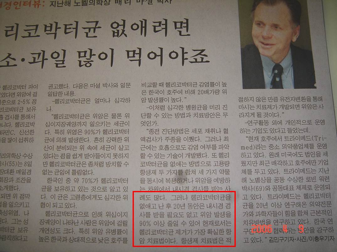
연구 결과를 보시면 득이 크지는 않습니다. 저는 약간 득이 될 것으로 봅니다. 그러나 제균 후 내시경 추적관찰을 게을리 할 정도로 제균의 득이 크지는 않습니다. False sense of safety를 주의해야 합니다. 과거 황당한 기분으로 읽었던 신문 기사를 소개합니다. "헬리코박터균을 없애고 난 후 20년 동안은 내시경 검사를 받을 필요도 없고 위암 발생을 90% 이상 줄일 수 있어..."라는 괴상한 주장입니다. 노벨상을 받았다는 사람이 한 말인데 어이가 없습니다. 요즘은 야쿠르트를 팔고 있으니 저는 별로 신뢰하지 않습니다. 문제는 국민들이 저같은 의사의 말보다 노벨상의 권위를 높게 생각한다는 점입니다. 노벨상이야 폭탄팔아 번 돈으로 만든 상일 뿐이데...
노벨상 수상자의 발언 중 최고 엉터리 발언입니다.임상의 문제를 명쾌하게 답해주는 좋은 연구는 흔치 않습니다. 연구는 잘 controlled 된 상태에서 이루어집니다. 환자는 제각각의 변수를 가지고 있습니다. 개별 의료진은 연구 결과와 배경을 잘 이해한 후 적절히 해석(혹은 extrapolation)해야 합니다. 의학을 예술이라 부르는 이유이기도 합니다. 철학이라 부르는 분도 계십니다.
[2017-3-3. 어떤 환자 질문]
내시경 검사 할 때마다 이상하다고 자꾸 조직검사를 합니다. 한번은 비정형 소견 (atypical)이 나와 큰 병원에서 재검까지 받기도 했습니다. 어떻게 안 될까요?
[2017-3-3. 이준행 답변]
헬리코박터를 제균하면 됩니다. 심평원에서 못하게 할 뿐입니다. 저는 제균치료를 권하였습니다. 심평원보다 환자의 건강과 행복이 중요하니까.
[2018-6-17. 이준행 수정 답변]
헬리코박터 제균치료를 권합니다. 2018년 1월 1일부터는 '약가 전액 환자 부담'으로 치료받으실 수 있습니다.
[2017-6-5 web-seminar 질문]
IDEN 2017 Ueo 선생님 강의 중 Kyoto system을 말씀하시면서 red streak는 Hp 음성이라고 하셨는데, 전 red streak를 chronic superficial gastritis로 받아들였습니다. 그런데, 오늘 chronic superficial gastritis의 대부분이 Hp 양성이라고 하셔서 좀 헷갈립니다. 두 설명이 상충되는 것 같습니다.
[2017-6-5. 이준행 답변]
매우 좋은 질문입니다. Chronic superficial gastritis의 정의가 명확하지 않기 때문에 발생한 혼선이라고 생각합니다.
Chronic superficial gastritis (CSG)는 아마도 위내시경의 아버지 Dr. Schindler가 처음 사용한 용어인 것 같습니다. 아래 그림과 같이 위축성 위염이나 비후성 위염이 아니면 모두 CSG입니다. Schindler의 CSG는 넓은 의미의 CSG이고 상당수가 Helicobacter 감염과 관련되어 있습니다.
일본에서 CSG는 조금 다른 방식으로 이해되고 있습니다. 2016년 내시경 세미나에서 소개된 바와 같이 일본에서의 CSG는 Hp 미감염자의 내시경 소견 (표재성 위염, 위저선 용종, 헤마틴) 중 하나로 간주되고 있습니다. 전정부에서 시작되는 선상의 발적을 말하며 위산 분비나 많은 상황에서 보이는 소견으로 이해됩니다.
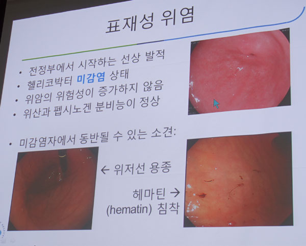
요컨데 우리가 흔히 말하는 CSG는 Schindler의 CSG, 즉 넓은 의미의 CSG로서 Hp 감염자가 많은 반면, 일본 Kyoto 분류에서 말하는 CSG는 좁은 의미의 CSG로서 Hp 미감염자의 특징적 소견입니다. 일본의 의학용어는 여타 나라와 다른 경우가 많습니다. 일본 문헌을 볼 때 매우 조심해야 합니다. 이러한 내용을 강의 중 명료하게 나누어 설명하지 못하여 죄송합니다. 아래 두 자료를 참고하시기 바랍니다.
* 참고: Sun-Young Lee. Endoscopic gastritis, serum pepsinogen assay, and Helicobacter pylori infection Korean J Intern Med 2016
* 참고 : 검진발견 소화성 궤양과 기타 위십이지장 질환 이선영. 2016년 내시경 세미나 강의
[2017-6-13. 애독자 질문]
... 설령 헬리코박터 위염이 맞다 그렇다 하더라도 r/o H. pylori gastritis라고 impression을 쓰는 것도 이상할 것 같습니다. 치료 indication도 아닌데, H. pylori 조직검사도 하지 않을 것이면서 r/o H. pylori gastritis 쓰는 것이 무슨 의미가 있을까 싶습니다. 환자가 '저건 왜 저렇게 빨간가요?'라고 묻는다면 뭐라고 대답해야할지... 그냥 '저 부분만 위염이 심한거에요' 이렇게 두루뭉술 넘어가야하는지...
[2017-6-13. 이준행 답변]
... 진단명이 문제인데요... "위체상부와 분문부 점막의 점상 반점, 미만성 발적 (diffuse redness)이라는 헬리코박터 위염의 특징적 소견을 보인 환자를 어떻게 기술할 것인지 고민입니다. 이런 환자는 제균치료를 하고 싶은데 대한민국 정부에서 허락하지 않고 있다는 점도 고민입니다. 일단 잠정적으로 chronic superficial gastritis of the proximal stomach라고 쓰고 있습니다. 물론 r/o Helicobacter-associated라고 붙이고 몇 개의 조직검사를 할 수도 있습니다. 안해도 무방합니다만..." 정도가 여전히 제가 생각할 수 있는 최선의 답입니다.
Strict한 급여 기준을 유지하면서 조금의 flixibility도 허용하지 않는 우리나라의 경직된 의료 현장에서 자주 만나는 안타까운 모습니다. 뭔가의 제도가 strict하게 되어 있으면 어딘가에서는 융통성을 허용해야 하는데 그 부분이 전혀 고려되지 않고 있습니다. 우리나라를 제외하고 off label 처방을 전혀 인정하지 않는 의료 시스템은 거의 없습니다. 그 비싼 미국 의료에서도 off label 치료는 인정됩니다. 일본에서는 누구나 검사하고 누구나 치료받는 헬리코박터 위염을 우리나라에서는 완전히 불법으로 간주하는 모습은 심평의학의 한계입니다. 적어도 원하는 사람이라도 치료를 받을 수 있게 해 주어야 합니다. 적어도 원하는 의사라도 삭감을 걱정하지 않고 처방할 수 있게 해 주어야 합니다. 우리나라 의학이 발전하려면, 우리 국민들이 제대로 된 의료 서비스를 받으려면 심평의학에서 벗어나야 할 것 같습니다. 박정희 독재정권 시대에 만들어지 권위주의 의료시스템이 수명을 다한 것 같습니다. 독재자의 딸도 대통령에서 물러난 마당이니... 이제 의료시스템도 제대로 바뀌어야 하지 않을까요.
[2018-6-17. 이준행 수정 답변]
진단명에 헬리코박터 위염이라고 쓰고 헬리코박터 검사를 하십시오 (90/100). 2018년 1월 1일부터는 '약가 전액 환자 부담'으로 제균치료를 할 수 있습니다.
[2018-10-31. 애독자 질문]
교수님 안녕하십니까? 엔도투데이로 공부하는 내과의사입니다.
Thickened gastric rugae with whitish, sticky exudate 는 acute H.pylori infection 이라고 엔도투데이로 배웠습니다. 가르쳐주셔서 감사합니다. 이런 소견이 있으면, 보만4형 위암을 감별하려고, 저는 위체부 대만에 조직검사를 할것 같은데요. 그리고 복부 CT 도 때로는 권고할것 같은데요. . 혹시나 모를 보만4형 놓치는 의료사고 대비하려는 방어적인 진료때문에... 교수님께서도 조직검사를 하시는지요? 제가 필요없는 조직검사를 하는가 싶어서, 과잉진료인가 싶어서, 교수님께 문의드립니다. 항상 감사드립니다.
[2018-10-31. 이준행 답변]
비록 헬리코박터 위염이 "Thickened gastric rugae with whitish, sticky exudate" 소견을 보이기는 하지만 보만 4형 진행성 위암과 사뭇 다른 모양입니다. Thickened fold에서는 항상 보만 4형 진행성 위암을 가장 먼저 고려해야 하겠습니다. 헬리코박터 위염이라고 가정하고 가볍게 보다가는 진단이 늦어질 수 있기 때문입니다. 조직검사는 언제나 과잉 진료가 아닙니다. 필요할 때에는 언제나 하면 됩니다. 남들이 다들 불필요하다고 하는 상황에서 나만 조직검사를 자꾸 필요하다고 생각하면 과잉입니다. 그런 분들 있습니다. 그렇더라도 내시경 하는 의사가 필요해서 하면 그 순간은 과잉이 아입니다. 말장난 같지만 그렇게 해석할 수 밖에 없습니다.
조직검사도 적당히 하는 것이 좋습니다. 우리나라는 일본에 비하여 대략 3배 정도로 많이 하고 있지 않나 생각합니다. 제 느낌입니다. 대충 보고 조직검사 많이 하는 것이 우리 나라 스타일이고 일본은 정반대입니다.
[2019-3-8] 위와 십이지장의 microbiota와 조직학적, 내시경적 염증 및 증상과의 관련성을 분석한 건국대 이선영 교수님 팀의 연구를 흥미롭게 보았습니다 (J. Clin. Med 2019).
저자께서는 "병리학적 위염과 내시경적 위염은 세균총의 구성이 유사하지만, 증상적 위염은 위내 세균총보다 십이지장 세균총이 더 연관되어 있고, 병리학적 위염이나 내시경적 위염과는 상이하다는 결론"으로 설명하고 계십니다.
Abstract: Mucosal inflammation is characterized by neutrophil and mononuclear cell infiltration. This study aimed to determine the gastric and duodenal microbiota associated with histological, endoscopic, and symptomatic gastritis. Dyspeptic adults who presented for evaluation were included. Subjects with either comorbidities or recent drug intake were excluded. Three endoscopic biopsies were obtained from the antrum, body, and duodenum. Next-generation sequencing for 16S ribosomal RNA V1-V2 hypervariable regions was performed. The correlation between the composition of microbiota and the degree of inflammatory cell infiltration, endoscopic findings, and Patient Assessment of Gastrointestinal Disorders Symptom Severity Index (PAGI-SYM) score was analyzed. In 98 included subjects, microbial communities in the antrum and body showed Bray-Curtis similarity; however, those in the duodenum showed dissimilarity. Histological and endoscopic gastritis was associated with the abundance of Helicobacter pylori and that of commensal bacteria in the stomach. The abundances of Variovorax paradoxus and Porphyromonas gingivalis were correlated with histological gastritis, but not with endoscopic or symptomatic gastritis. The total PAGI-SYM score showed a stronger correlation with the duodenal microbiota (Prevotella nanceiensis and Alloprevotella rava) than with the gastric microbiota (H. pylori, Neisseria elongate, and Corynebacterium segmentosum). Different correlations of the gastric and duodenal microbiota with histological, endoscopic, and symptomatic gastritis were observed for the first time at the species level. H. pylori-negative gastritis is not associated with endoscopic or symptomatic gastritis. Only H. pylori-induced endoscopic gastritis requires gastric cancer surveillance. Owing to the weak correlation with H. pylori, symptomatic gastritis should be assessed separately from histological and endoscopic gastritis.
Figure 3. Significant endoscopic findings that were correlated with the relative abundance of microbiota. Helicobacter pylori was abundant in the presence of hemorrhagic spots, hypertrophic rugae, advanced atrophy, and mucosal nodularity, whereas Pseudomonas veronii and Propionibacterium acnes were abundant in the absence of these findings. Pseudomonas sp. was abundant in the absence of atrophy and nodularity. Moreover, Cloacibacterium rupense and Staphylococcus epidermidis were abundant in the absence of nodularity. The median relative abundance of each species were provided with range (minimum-maximum) using the Kruskal-Wallis test. H. pylori: Helicobacter pylori; P. veronii: Pseudomonas veronii; P. acnes: Propionibacterium acnes; C. rupense: Cloacibacterium rupense; S. epidermidis: Staphylococcus epidermidis.
이 분야 연구 경험이 없는 저로서는 논문의 세부 내용을 모두 이해하기는 어려웠습니다만 흥미로운 자료는 많았습니다.
- Hp는 조직학적 위염이나 내시경적 위염과는 관련되어 있었으나 증상과는 관련성이 없었습니다.
- Hp 이외의 몇 세균이 조직학적 위염과 관련되어 있었으나 내시경적 위염이나 증상을 유발하지는 못했습니다. The abundances of Variovorax paradoxus and Porphyromonas gingivalis were correlated with histological gastritis, but not with endoscopic or symptomatic gastritis.
- 위장관 증상은 십이지장 세균총과 관련성을 보였습니다. The total PAGI-SYM score showed a stronger correlation with the duodenal microbiota (Prevotella nanceiensis and Alloprevotella rava) than with the gastric microbiota (H. pylori, Neisseria elongate, and Corynebacterium segmentosum).
- Hp에 감염되면 위 세균총의 다양성이 감소되면서 (전정부보다 위체부의 다양성이 더 감소), 동시에 십이지장 세균총의 다양성은 증가되었습니다. H. pylori-infected subjects showed lower diversity indices in the stomach, and higher diversity indices in the duodenum than non-infected subjects. (제가 기전을 이해하기 어려운 부분이었습니다.)
연구자가 본인의 외래를 방문한 환자를 대상으로 연구 참여를 권유하고 동의서를 받고 증상 설문과 검체를 얻어 진행된 연구였습니다. 흥미로운 언급이 있었습니다.^^ "Most of the patients who visited the clinic were female subjects who wanted to be examined by a female gastroenterologist; thus, only 21 of out included 98 subjects were male."
저자의 결론을 옮깁니다. 결론의 마지막 문장이 정확히 어떤 의미인지 궁금했습니다.
Different correlations of the gastric and duodenal microbiota with histological, endoscopic, and symptomatic gastritis were observed for the first time at the species level. Histological gastritis was associated with the relative abundances of H. pylori, V. paradoxus, and P. gingivalis. H. pylori-negative gastritis was not associated with endoscopic or symptomatic gastritis. H. pylori was the only pathogen associated with endoscopic gastritis, which requires gastric cancer surveillance.
Symptomatic gastritis should be evaluated and managed differently from histological and endoscopic gastritis, because it is more strongly correlated with the duodenal microbiota (P. nanceiensis and A. rava) than the gastric microbiota (H. pylori, N. elongata, and C. segmentosum). Thus, factors other than H. pylori infection status should be assessed in cases of symptomatic gastritis.
무척 힘든 연구를 끝까지 마치고 멋진 모습으로 발표하신 연구자들에게 격려과 축하의 메세지를 보냅니다. 고생하셨습니다.
[이선영 교수님 답변]
증상적 위염이 비록 위세균총보다 십이지장 세균총과 연관이 있다고 나왔지만, 세균총 이외의 요인이 증상 발현과 연관이 있을 거라 생각합니다. 리뷰어들은 세균총만 강조하라 했지만, 젊은 여성에서 위염 증상이 흔한 걸 보면 성별, 나이, 매운 음식, 예민한 성격 등이 증상 발현에 있어 세균총보다 더 중요하다고 생각합니다.
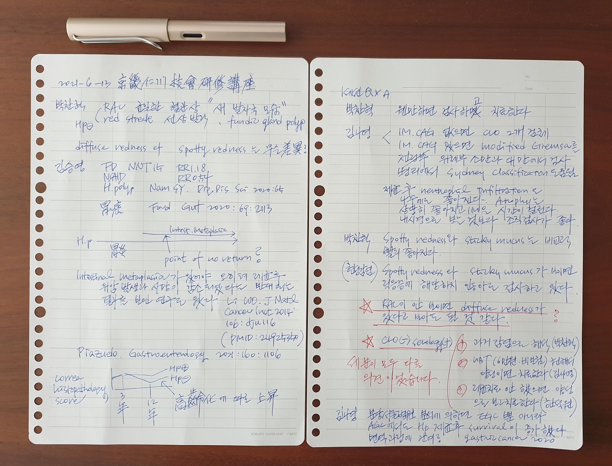
[2021-6-13. 내시경학회 경인지회 심포지엄 헬리코박터 세션을 듣고 이준행 자문자답]
질문: 헬리코박터 위염 중 diffuse redness와 spotty redness는 어떻게 다릅니까?
답변: 일본의 Kyoto classification 책자에는 둘이 구분되어 있습니다. 책자에 나온 정의와 sample 사진은 아래와 같습니다.
- Diffuse redness refers to uniformly reddish mucosa with continous expansion observed in non-atrophic mucosa main in the body.
영문판 Kyoto Classification of Gastritis 38쪽
- Spotty redness is marked by irregular red dots of various shapes and sizes.
영문판 Kyoto Classification of Gastritis 43쪽
우리나라에서는 diffuse redness와 spotty redness를 섞어서 모두 diffuse redness로 부르는 경향이 있습니다. EndoTODAY에서도 그러한 입장입니다. 이유는 일본에서 말하는 diffuse redness는 너무 주관적이고 기계의 minor setting에 영향을 받으므로 reproducibility가 떨어지기 때문입니다.
2021년 6월 13일 경인지회연수강좌에서 박찬혁 교수님은 둘을 구분하여 설명하셨습니다. 따라서 그에 대한 질문이 있었는데 "RAC이 안 보이면 diffuse redness가 있다고 보아도 될 것 같다"는 답변이 나왔습니다. 저는 더 혼란스러워졌습니다. 다음 세션 연자께서도 이 부분을 comment하셨는데 막상 diffuse redness라는 제목의 slide에서는 spotty redness 소견이었습니다. 역시 우리나라에서는 대부분 이 둘을 구분하지 않는다고 보는 것이 좋겠습니다. 사실 일본에서 모든 사람이 이 둘을 정확히 구분하고 있는지 미지수입니다. 몇 분의 일본 선생님들 강의를 들어보아도 diffuse redness와 spotty redness를 잘 구분하지 않는 것 같았기 때문입니다.
Kyoto 분류를 처음 제안한 팀에서는 둘을 구분하고자 하였으나 일본이나 우리나라의 내시경실 현장에서는 둘을 구분하는데 어려움을 겪고 있고 결국 마구 섞어 쓰고 있다고 보는 것이 정확할 것 같습니다.
[2021-6-16. 건국대 이선영 교수님 comment]
건국대 이선영 교수님께서 EndoTODAY 블로그 포스팅을 보시고 아래와 같은 의견을 보내주셨습니다.
위염의 내시경소견 책에서 설명하는 걸 빠뜨렸던 부분이라 늘 마음에 걸렸었는데, 어제 마침 diffuse vs. spotty redness에 대한 질문을 올리셔서 말씀드립니다.
첨부화일 3쪽의 사진 ab와 같이 diffuse redness는 위 전체에서 광범위하게 관찰될 수 있는 미세한 발적이고, spotty redness는 diffuse redness 중에서 사람 눈에 보일 정도로 커진 발적들이 dot나 petechiae처럼 보이는 것이라고 합니다.
Spotty redness는 두께가 얇은 기저부에서 주로 관찰되기 때문에 감염자 감별 시 fundus 부근만 관찰하면 알 수 있지만, diffuse redness는 위 전체에 걸쳐 퍼진 미세한 발적이기 자세히 봐야만 알 수 있습니다. 즉, 내시경 검사 시 한 눈에 보이는 기저부의 hemorrhagic spots은 spotty redness이며, spotty redness 바탕에 깔린 위 전체의 미세한 발적은 diffuse redness입니다. 자세히 봐야만 알 수 있는 미세한 혈관확장들인 셈입니다.
감염자는 대부분 둘 다 있어서 저는 diffuse spotty redness라고 표현하기도 합니다.
[2022-4-16 SIDDS 이선영 교수님 강의]
[2022-5-3. 알림]
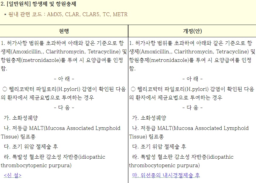
2022년 5월 1일로 '항생제 및 항원충제' 등 약제의 고시가 일부 변경되었습니다. 위선종의 내시경절제술 후 헬리코박터 파일로리(H.pylori) 감염이 확인된 경우 기존 전액 환자 부담에서 급여로 변경되었습니다.
[2025-1-26. 애독자 질문]
Alcoholics case
[2025-1-26. 이준행 답변]
좋은 질문 감사합니다. 증례에서는 일단 지혈술 후 즉시 헬리코박터 검사를 하였습니다. 당시는 Giemsa 음성이었습니다. 물론 출혈 상황에서 헬리코박터 조직검사의 양성률이 조금 낮을 수 있다는 점은 이해하고 있습니다. Helicobacter 양성일 것으로 예상되는 상황에서 음성이 나왔으므로 추적검사에서도 헬리코박터 검사를 하는 것이 좋은 상황이었습니다. PPI를 4주 처방하고 3주 후 내시경 검사를 하였으므로 질문주신 바와 같이 PPI를 유지한 상태로 내시경 재검을 한 것은 맞습니다.
저도 소화성궤양 환자의 추적검사에서 PPI를 2주 중단하고 내시경을 하면서 헬리코박터 검사를 합니다. 그러나 고위험 출혈 환자에서, 특히 만성 질환이 있거나 NSAIDs나 술을 끊지 않은 환자에서 PPI 중단 후 내시경 검사를 하기 전 1-2주 사이에 재출혈을 하는 경우를 경험한 바 있습니다. 이런 위험이 있는 경우에는 헬리코박터 검사의 정확도가 다소 낮아지는 것을 감수하더라도 on PPI 상태에서 내시경 검사를 하고 있습니다. 헬리코박터는 추후 언제든지 검사할 수 있기 때문입니다. "이러저러한 이유로 헬리코박터 검사를 충분히 할 수 없었으니 이제 헬리코박터 검사를 해 봅시다"라고 말할 기회는 많습니다. UBT를 하면 됩니다. 헬리코박터 제균치료 후라면 일반 보험 급여, 헬리코박터 제균치료를 하지 않았던 환자에서는 비급여입니다.
![]() [2025-2-10] 이선영 교수님의 위염 강의에서 많은 것을 배우고 있습니다. 오늘의 강의는 정말 좋았습니다. Wonderful
[2025-2-10] 이선영 교수님의 위염 강의에서 많은 것을 배우고 있습니다. 오늘의 강의는 정말 좋았습니다. Wonderful
![]() [2025-10-25/26] 추계학술대회 및 헬리코박터 아카데미
[2025-10-25/26] 추계학술대회 및 헬리코박터 아카데미


태정현 교수님
태정현 교수님
태정현 교수님
임신 시 피해야 할 약
임신 시 피해야 할 약
보험제도
헬리코박터 위염
헬리코박터 위염
자자면역위염
![]() [References]
[References]
1) EndoTODAY 위염
2) 위염의 다양한 내시경 소견 및 그 의미 이선영 교수님. PDF
3) 이선영 교수님의 위염 관련 저술과 강의
- 리뷰 article - Endoscopic gastritis, serum pepsinogen assay, and Helicobacter pylori infection Korean J Intern Med 2016
- HUG 2025 강의 log-in 要
4) EndoTODAY 초보자를 위한 one point lesson - 위염
5) 無答토론 - 만성위염 내시경진단 003 2015년 헬리코박터 위염에 대한 관심이 부족하던 시절의 질문과 답변입니다.
6) 에라 모르겠다. Thickened fold by Helicobacter pylori gastritis
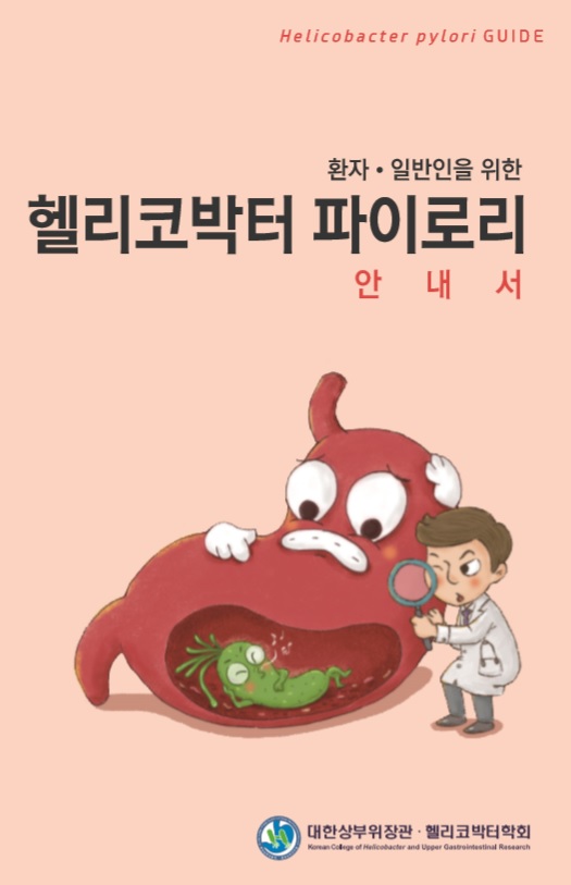
7) 2018-12 환자, 일반인을 위한 헬리코박터 파이로리 안내서
8) Endoscopic features of H. pylori infection 주문경 (VOD, IDEN2021)
9) 헬리코박터 위염을 내시경으로 발견하기 대한소화기내시경학회 KSGE webinar 2023/12 (log-in required)
10) 헬리코박터 - 일반 시민을 위한 9가지 핵심 질문과 답변 PDF (2026)
© 일원내시경교실 바른내시경연구소 이준행. EndoTODAY Endoscopy Learning Center. Lee Jun Haeng.
