EndoTODAY 내시경 교실
Beginner | ESA | Schedule | OPD
Seminars | Atlas | Recent | Links
 [StomachTODAY 051. Inflammatory myofibroblastic tumor (IMT)]
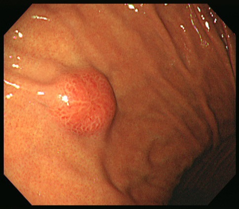
건강검진 내시경에서 발견된 점막하종양으로 의뢰된 분이다. 크기는 15mm 정도로 추정되었으며 표면은 발적되어 있었으며 4 방향에서 bridging fold가 모이는 양상이었고 조직검사에서는 특이소견이 없었다. 발열이나 체중감소, 피로감 등 전신증세는 없었다.
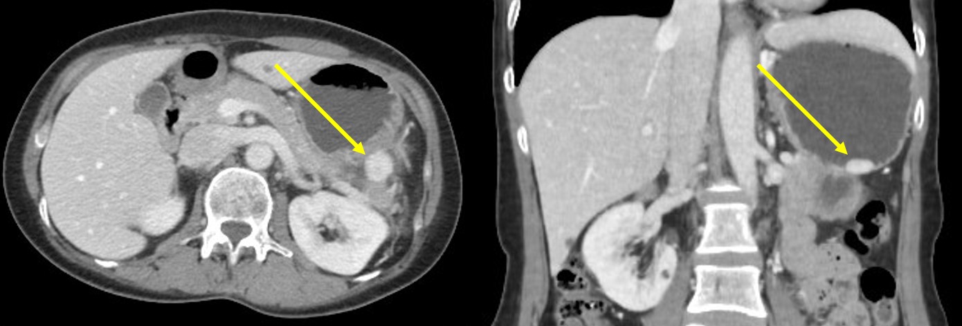
CT를 시행하였다. 위체상부 대만 후벽측에 1.9 cm까지 측정되는 hyperdense한 난형 병소가 관찰되었고 GIST 혹은 다른 종류의 혈관과다종괴로 추정되었다.
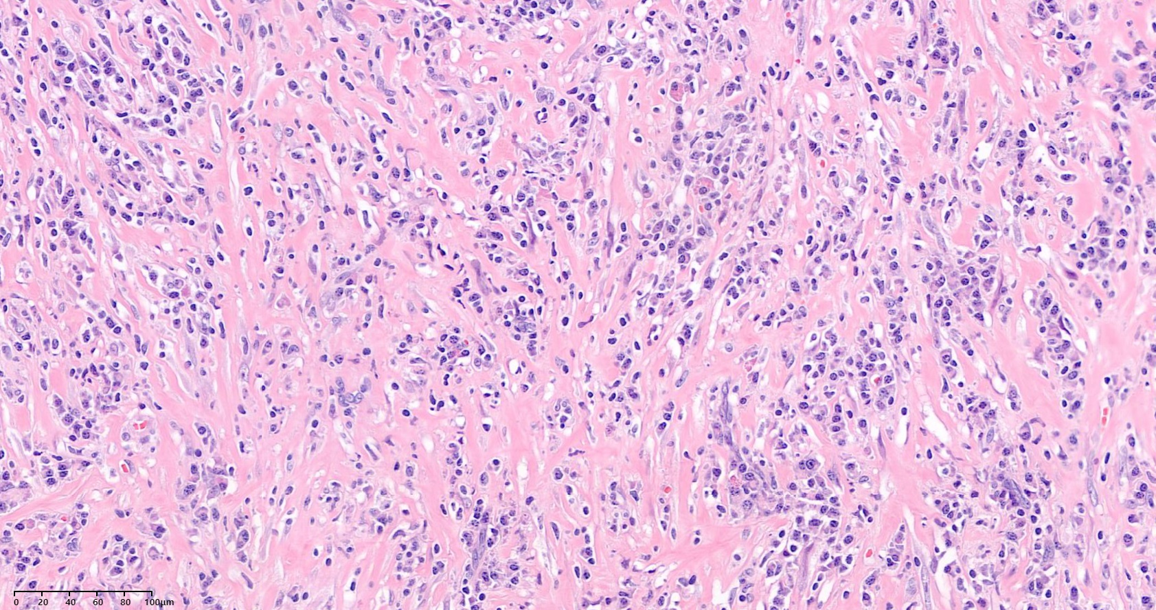
복강경 쐐기 절제술이 시행되었다. 병리 소견에서 방추형 세포가 소용돌이 모양을 이루고 있고 염증세포의 침윤이 동반되어 있었다. Actin 양성, CD23 음성, ALL 음성, Kappa와 Lambda는 polyclonal하게 양성이었다. 염증성 근섬유모세포성 종양 (Inflammatory myofibroblastic tumor IMT)으로 진단되었다. 1년 후 추적검사에서 재발 소견이 없었다.
염증성 근섬유모세포성 종양 (IMT)는 과거에는 반응성 변화로 생각하여 inflammatory myofibrohistiocytic proliferation, inflammatory pseudotumor 등으로 불리웠으나 이제는 종양성으로 간주되어 IMT로 불리는 종양이다. 소아, HIV 감염, 만성 육아종성 질환에서 흔하며 EBV나 HHV-8 감염과 관련된다는 주장이 있다. 조직학적으로 근섬유모세포의 증식과 형질세포, 림프구 및 조직구가 함께 출현한다. ALK gene rearrangement가 50%에서 발견된다. 폐에 발생하는 경우가 가장 많으며 복부, 후복막, 안와, 경부, 중추신경계 등 어디에서나 발생할 수 있다. 위장관에서는 복통, 복부 종괴, 설사, 장폐색 등의 증상을 일으킬 수 있다. 발열, night sweat, 체중감소, 피로감 등의 전신 증상을 동반할 수 있다. 우리나라에서는 무증상 성인에서 발견될 수 있다. 수술적 절제가 원칙이며 ¼에서 국소재발을 하며 드물게 전이를 보일 수 있다. 국소재발 병소에 대해서는 추가적인 절제술을 시행한다.
[More cases]
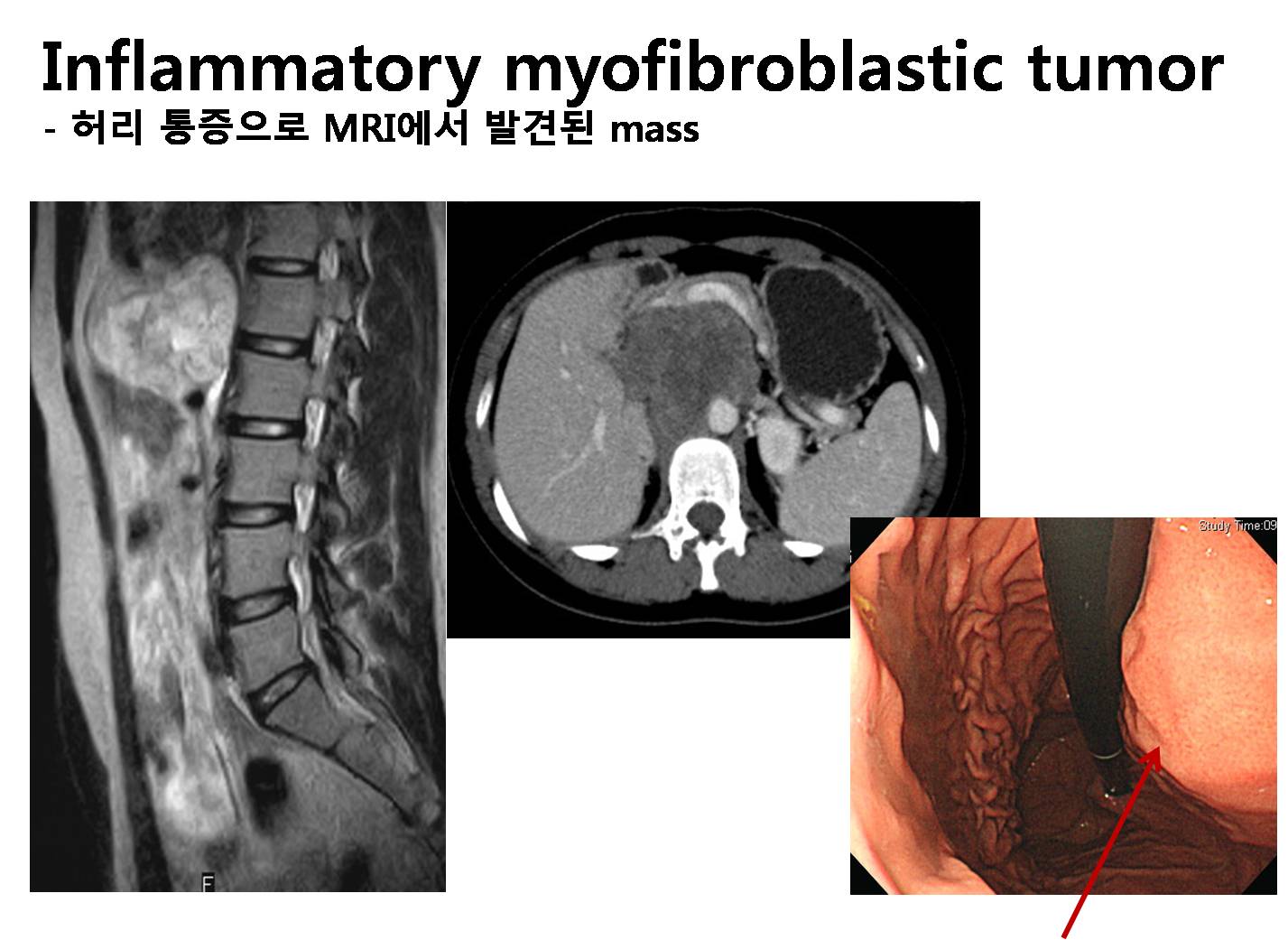
20대 여성이 허리 통증으로 시행한 spine MRI에서 mass가 발견되었습니다 (MRI 판독: L1-2 level의 aortocaval area로 7.7 x 6.3 cm의 solid mass로 생각되는 retroperitoneal mass). 저는 lymphoma의 가능성이 크지 않나 생각하였지만 stomach CT에서는 “Primary retroperitoneal mass such as neurogenic tumor, more likely”라는 impression과 함께 mass의 T2 signal이 높고 불균질하며 내부에 calcification이 있는 점은 전형적인 lymphoma와는 맞지 않는 소견이라는 의견을 얻었습니다. 내시경에서는 위체부의 extrinsic compression으로 관찰되었습니다. 수술을 시행하였고 inflammatory myofibroblastic tumor (IMT)라는 최종 진단을 받았습니다. IMT는 intermediate biologic potential을 가지는 neoplasm으로 참고 문헌의 일부를 소개한다.
Coffin CM, et al. Semin Diagn Pathol 1998;15:85-101. IMT or inflammatory pseudotumor was initially recognized in the lung, and somewhat later, a similar-appearing pathological process was reported in the liver. Presently, this tumor has been described in virtually all major organs and extrapulmonary sites with a few exceptions. It was thought initially that the IMT was nonneoplastic and represented an aberrant inflammatory response despite its gross and microscopic features of a spindle cell neoplasm. The inflammatory hypothesis about the pathogenesis has been more readily accommodated in the lung than in the extrapulmonary sites of involvement. Some cases, however, were accompanied by the constitutional symptoms and signs of an inflammatory process, which resolved in most cases after surgical resection. There were some pathological aspects of the IMT that seemingly contradicted its purely inflammatory nature, including its potential for local recurrence; development of multifocal, noncontiguous tumors; infiltrative local growth; vascular invasion; and malignant transformation. These pathological features seemed to support the hypothesis that the IMT is a neoplastic process, which has been augmented by reports that these tumors have clonal characteristics. Because these tumors have a predilection for children, embryonal rhabdomyosarcoma is another diagnostic temptation when an IMT presents in the bladder or other hollow viscus.
* 참고: EndoTODAY inflammatory pseudotumor
 [References]
1) EsoTODAY - 십이지장질환 증례토의
2) SmallTODAY - 소장질환 증례토의
3) ColonTODAY - 대장질환 증례토의
4) Dr. Sinn's LiverTODAY - 간질환 증례토의
5) 십이지장에서 발생한 염증성 근섬유모세포종 1예 Korean J Gastroenterol 2018;72:28-32 (부산대 증례)
© 일원내시경교실 바른내시경연구소 이준행. EndoTODAY Endoscopy Learning Center. Lee Jun Haeng (2022-7-30, update: 2024-8-16)