EndoTODAY 내시경 교실
Beginner | ESA | Schedule | OPD
Seminars | Atlas | Recent | Links
![]() [Schwannoma. 신경초종] - 終
[Schwannoma. 신경초종] - 終
1. Introduction
5. More cases of gastric schwannoma
9. References
![]() 1. Introduction to schwannoma
1. Introduction to schwannoma
Primary gastrontestinal neurogenic tumors are rare. They fall into two major groups: Those of peripheral nerve sheath origin (schwannomas, neurofibromas, ganglioneuromas, neuromas, and perineuromas) and those arising from sympathetic or chromaffin system (neuroblastomas, ganglioneuromas, and paragangliomas). Most schwannomas arise from the myenteric plexus.
Schwannoma (신경초종)는 Schwann 세포 기원하는 양성 종양으로 호발부위는 vestibular area (cerebellopontine angle)이고 neurofibromatosis type 2와 연관되어 있습니다. 위 schwannoma는 schwannoma의 위장관 침범 중에는 가장 흔하며 무증상 혹은 출혈이나 복통으로 발견되기도 합니다.
Stomach, mid body, anterior wall, wedge resection : Schwannoma
Actin (Smooth muscle) : Negative in tumor cells
Desmin : Negative in tumor cells
DOG-1 : Negative in tumor cells
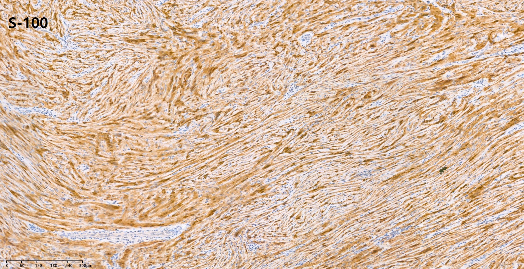
S-100 : Positive in tumor cells
Ki-67 : Positive in 5-10% of tumor cells
C-KIT (CD 117) : Negative in tumor cells
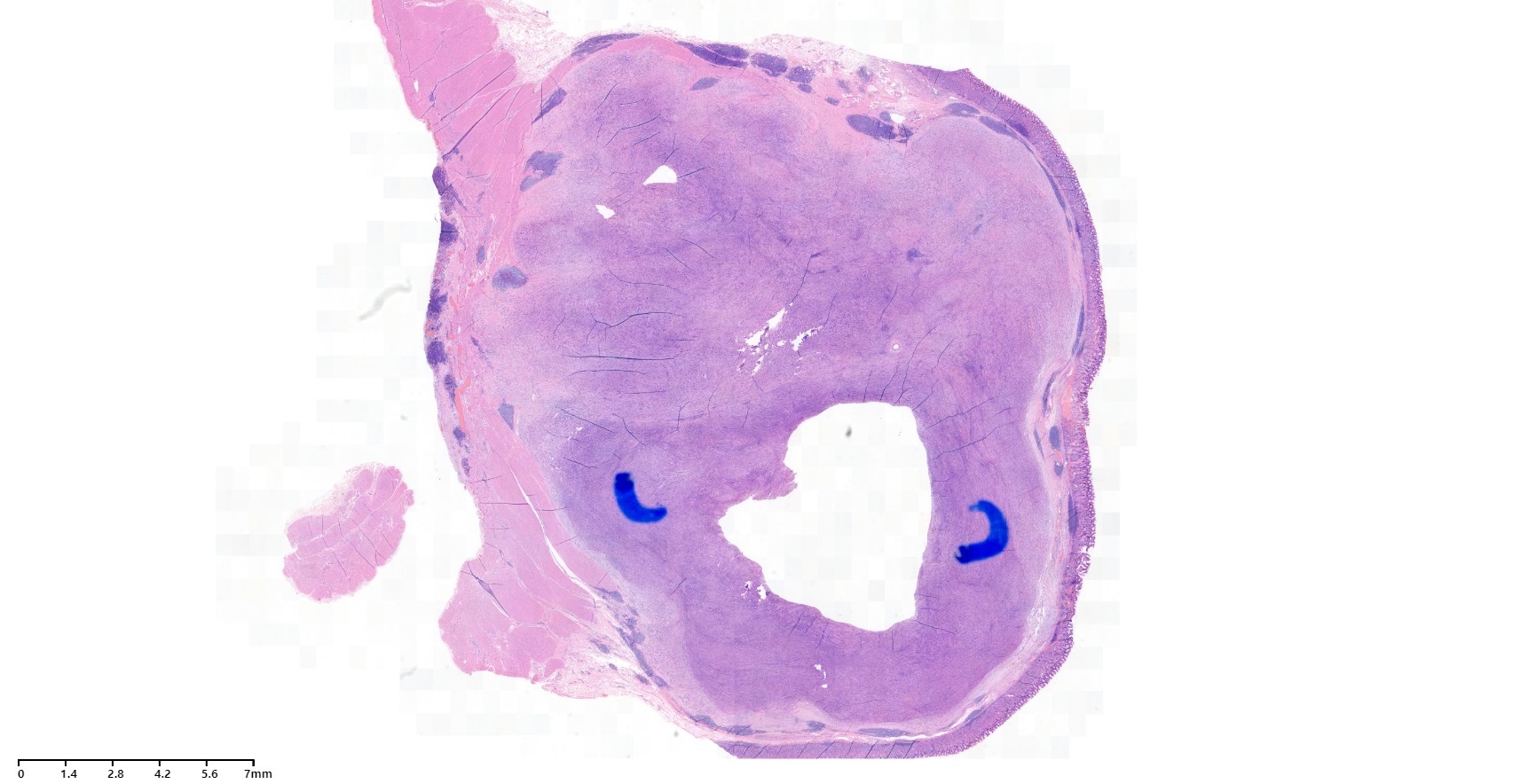
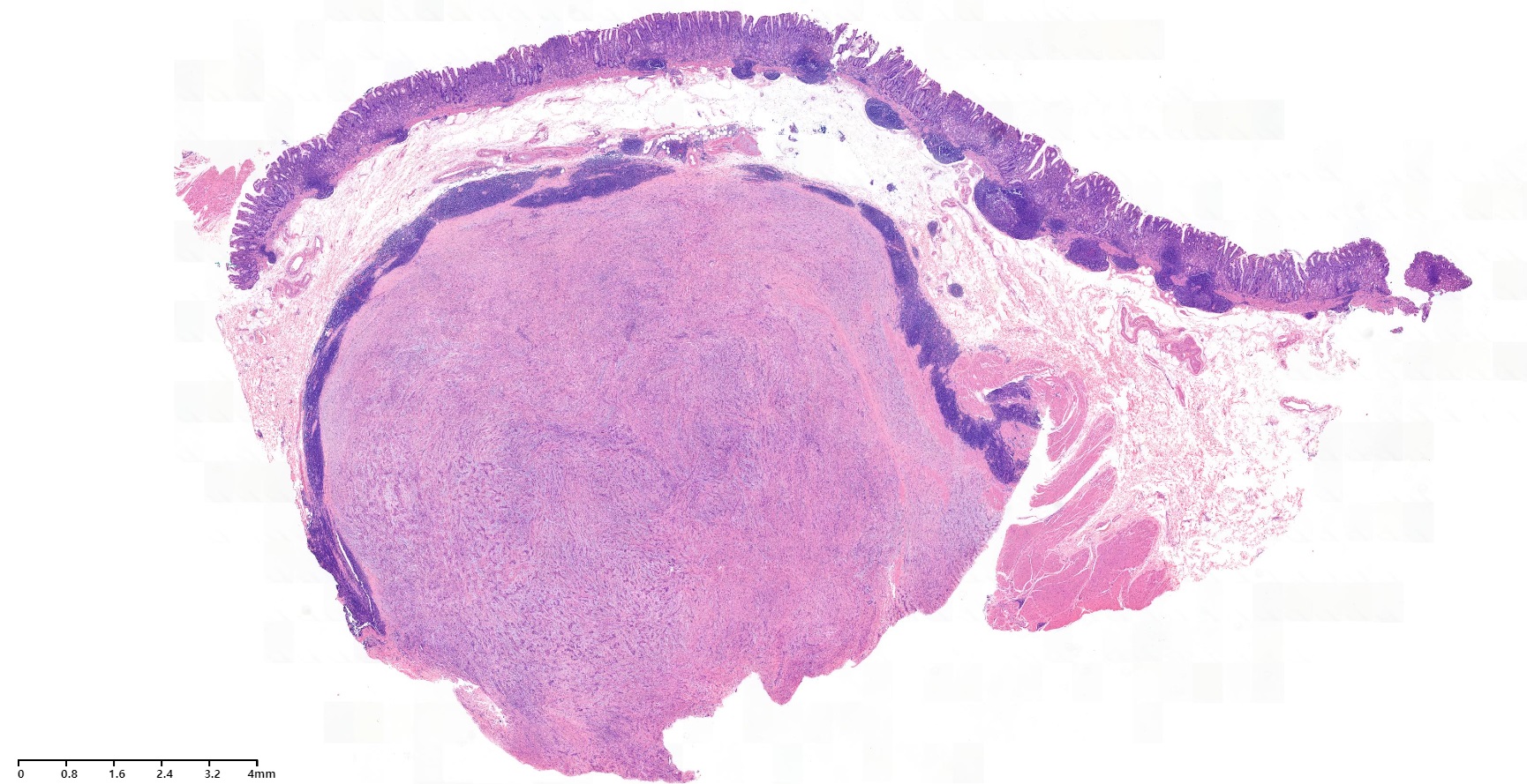
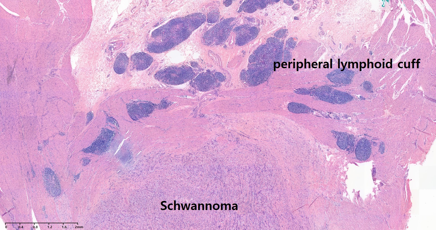
GI tract의 신경초종은 prominent lymphocytic rim을 보일 수 있습니다.
S-100
또 다른 증례입니다.
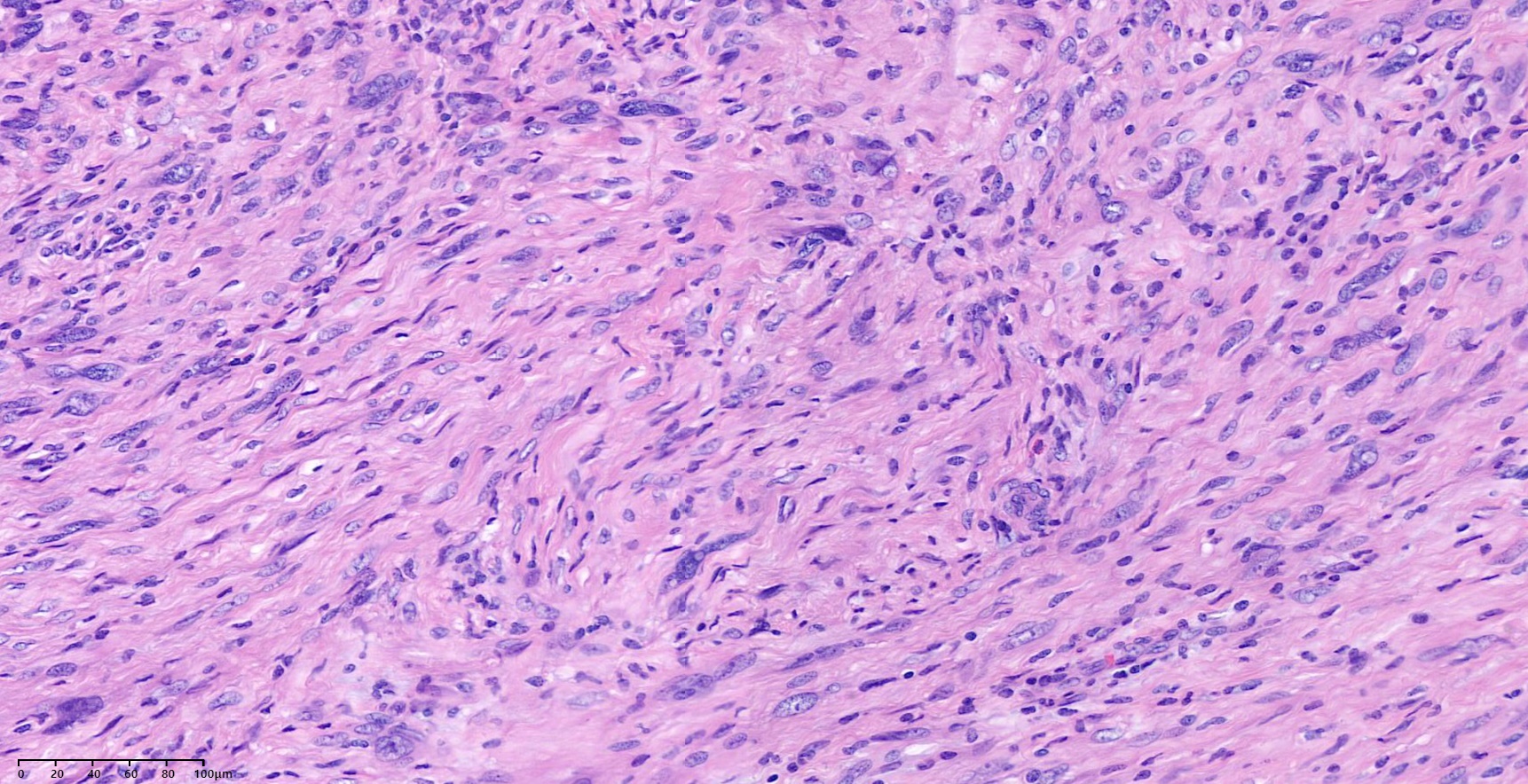
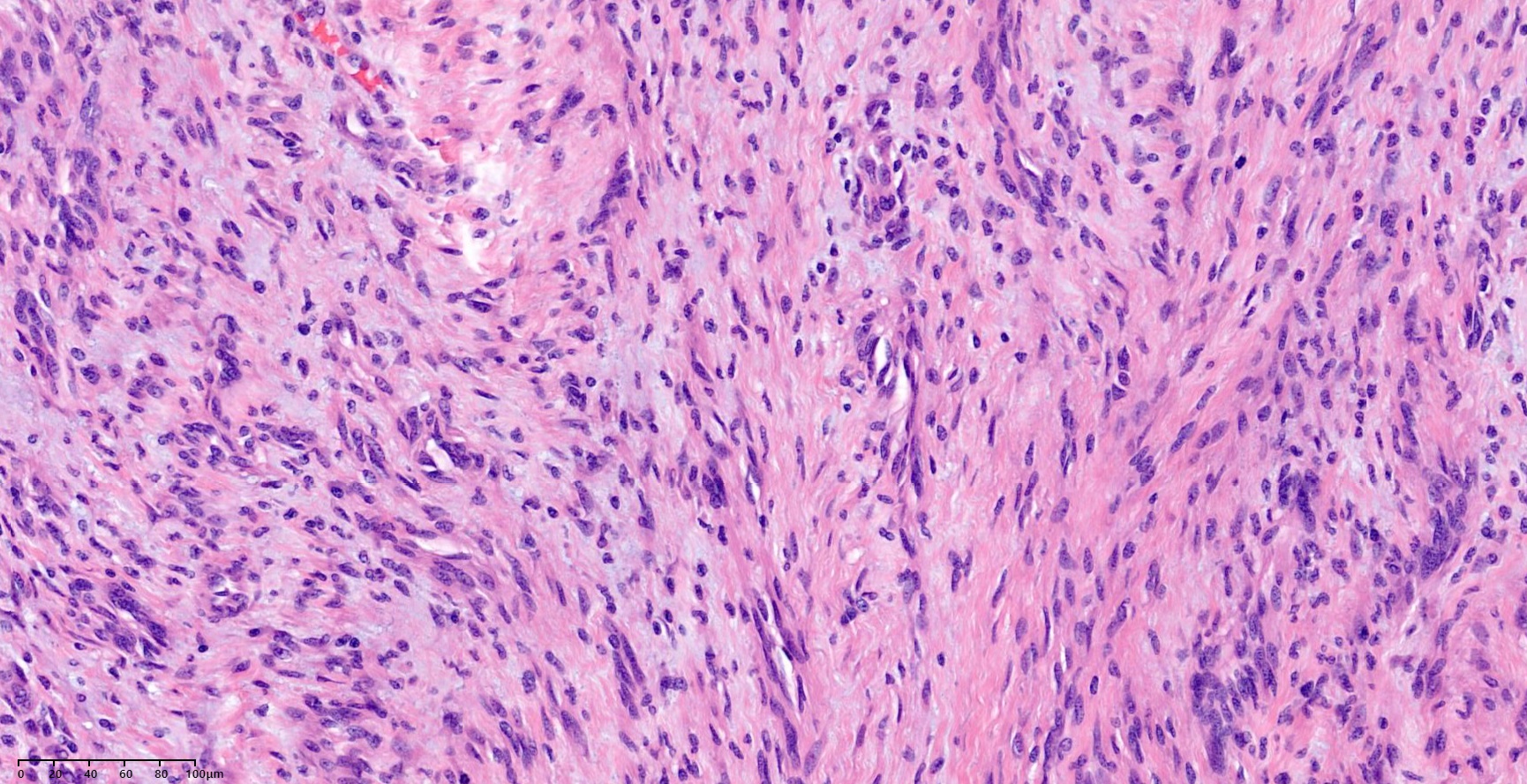
Benign schwannomas consist almost entirely of woven nests of compact bundles of slender, wavy S100-positive tumor cells with a loose myxoid stroma. The tumor cells may also form compact fascicles, occasionally aligning in rows or vague palisades (Verocay bodies). Antoni A area: interlacing bundles of spindle cells with wavy or oval nuclei eosinophilic cytoplasm, indistinct cytoplasmic borders. Antoni B area: hypocellular area
S-100
호발연령은 30-50세이며 호발부위는 이상하게도 lesser curvature쪽이라고 합니다. Intramural type이 가장 흔하지만 extraluminal type 혹은 endoluminal type도 가능합니다. 전형적인 형태는 궤양을 동반한 점막하 종양이며 악성변화는 매우 드물다고 합니다. 병소 주변에 lymph node가 커진 경우가 많은 것이 특징입니다. 따라서 benign인데도 수술로 연결되는 경우가 많습니다. 국소 절제술로 치료합니다.
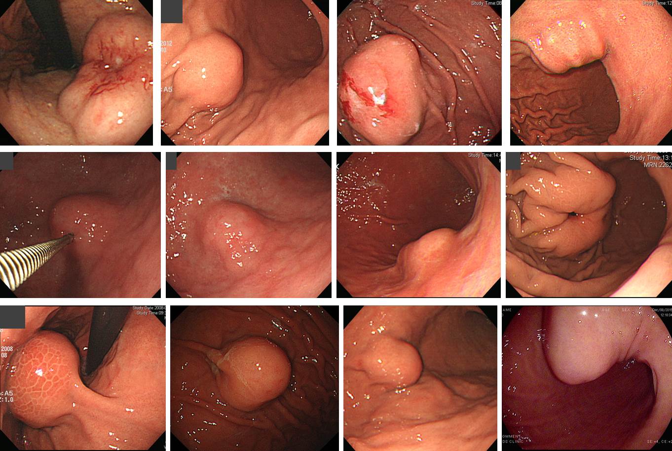
아래는 gastric schwannoma 증례 모음입니다. 다양한 소견을 보실 수 있습니다.
![]() 2. Endoluminal type
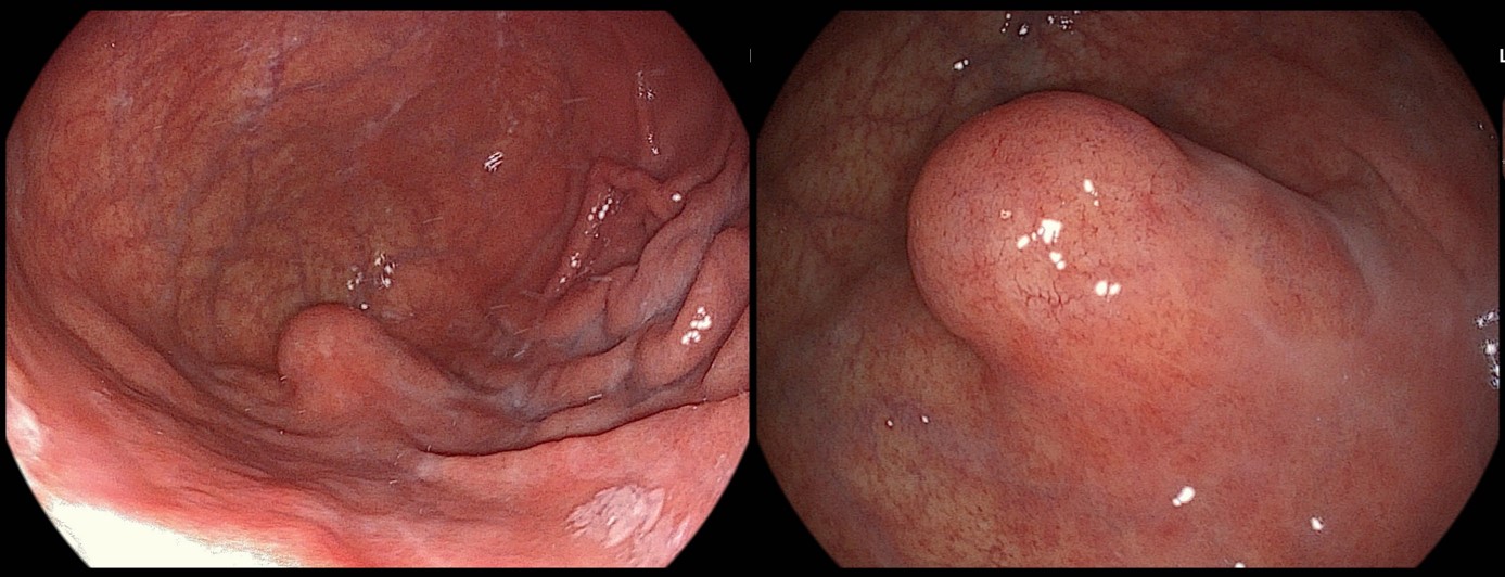
2. Endoluminal type
50대 여성에서 발견된 위 SMT로 수술을 시행하였고 최종 병리결과는 Schwannoma, 4.7 x 4.3 cm, negative resection margins, S-100: Positive였습니다.
4.5 x 4 x 3 cm schwanomma
![]() 3. Intramural type
3. Intramural type
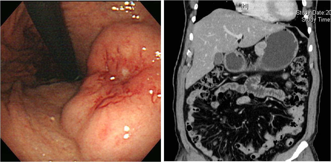
![]() 4. Extraluminal type
4. Extraluminal type
건강검진에서 우연히 발견된 SMT가 점차 커진다고 의뢰된 분으로 수술로 절제하였습니다.
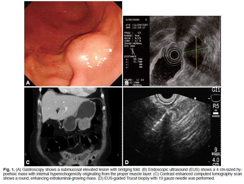
[2014-11-2. 추가] Clinical Endoscopy 2013년 5월호에 gastric schwannoma 증례가 실렸습니다 (link). EUS-guided trucut biopsy로 수술전 진단을 할 수 있었던 증례입니다.
이 정도 크기의 SMT라면 어짜피 wedge resection를 해야 합니다. 수술 전에 EUS-guided trucut biopsy가 꼭 필요했는지 약간 의심입니다.
![]() 5. More cases of gastric schwannoma
5. More cases of gastric schwannoma
Schwannoma (5.5 x 5 cm)
Schwannoma (2 x 1.3 cm), C-kit: Negative, S-100: Positive in tumor, SMA: Focal positive, in probably entrapped smooth muscle, CD34: Negative
Schwannoma (3.3 x 2.8 x 2.7 cm)
Schwannoma (2.3 x 1.5 cm)
Schwannoma (2.9 x 1.8 cm)
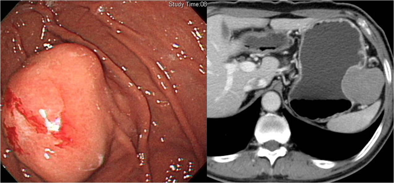
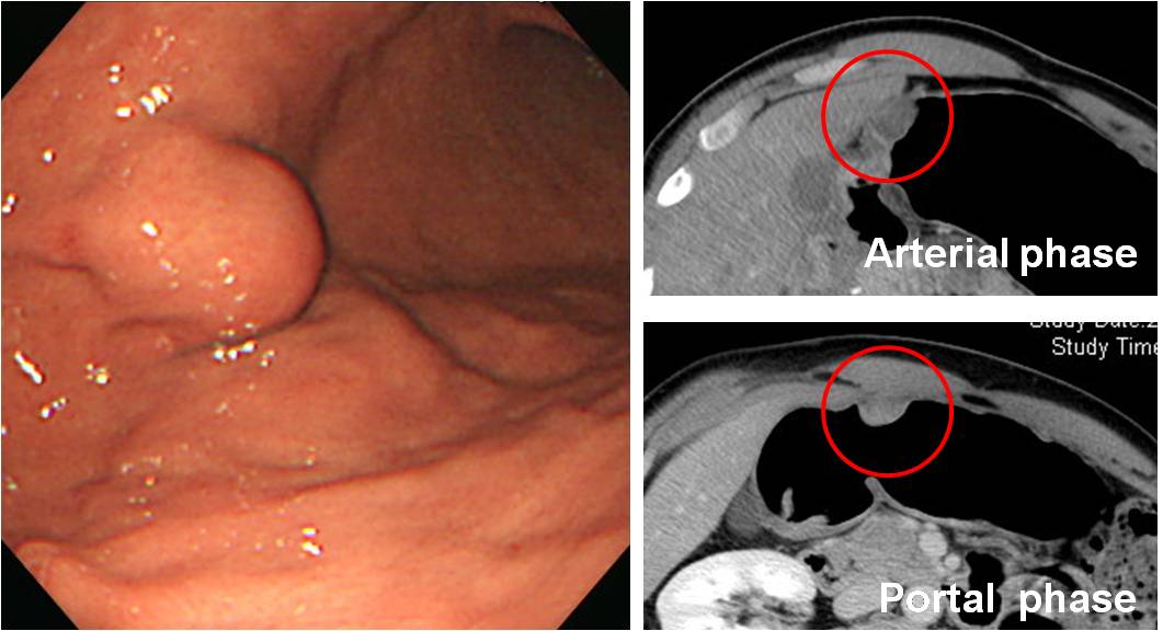
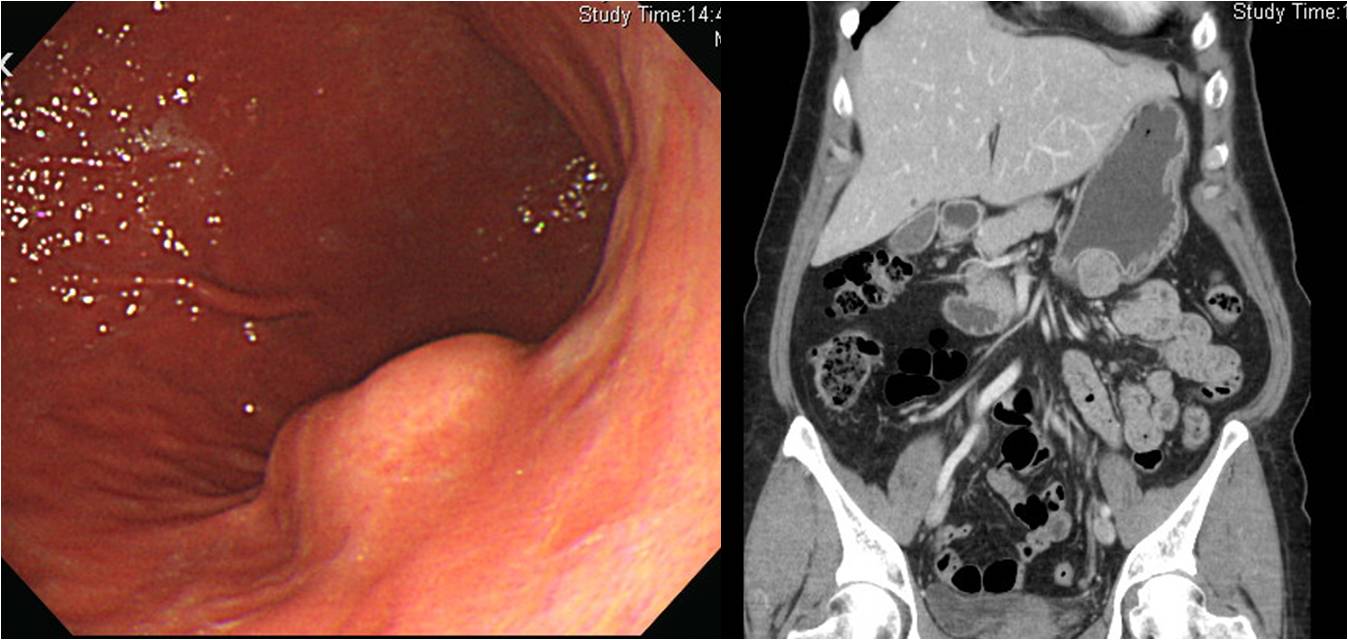
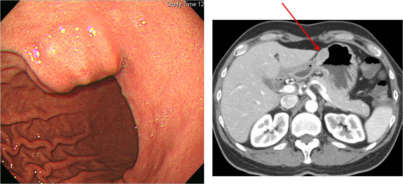
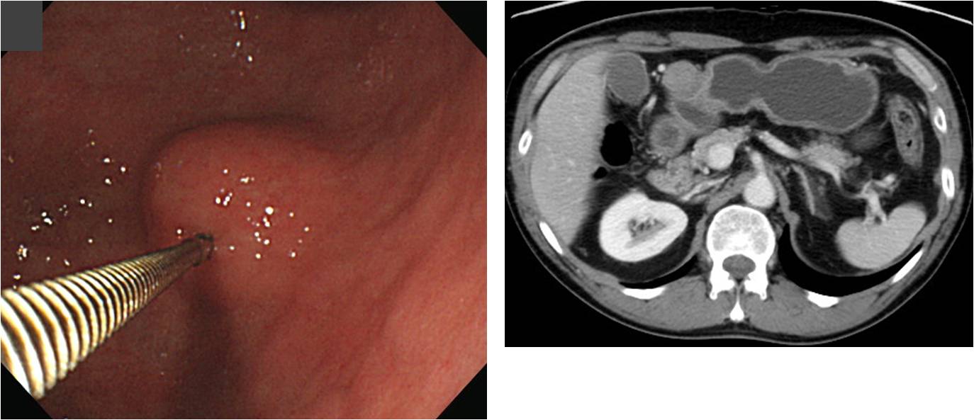
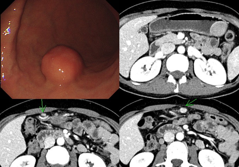
![]() 6. CT findings of schwannnoma
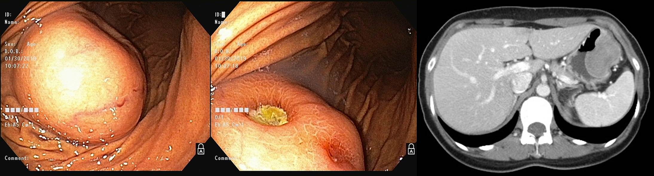
6. CT findings of schwannnoma
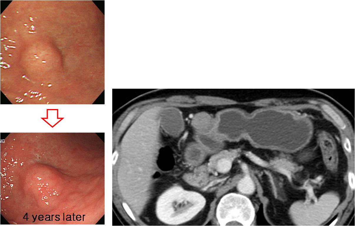
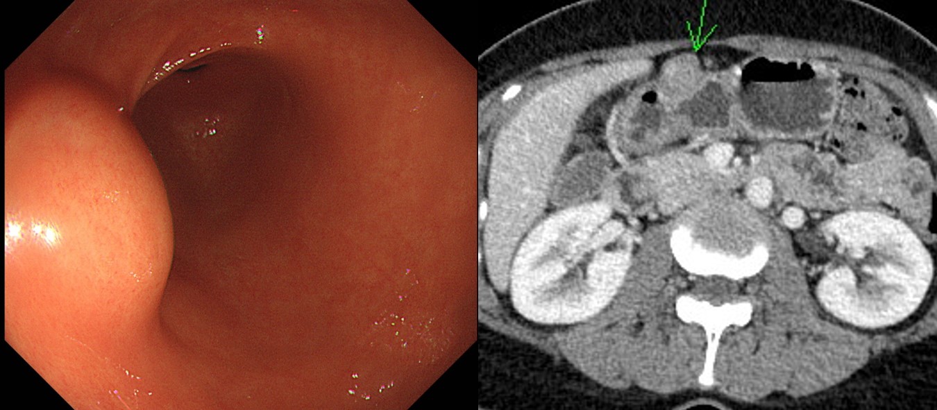
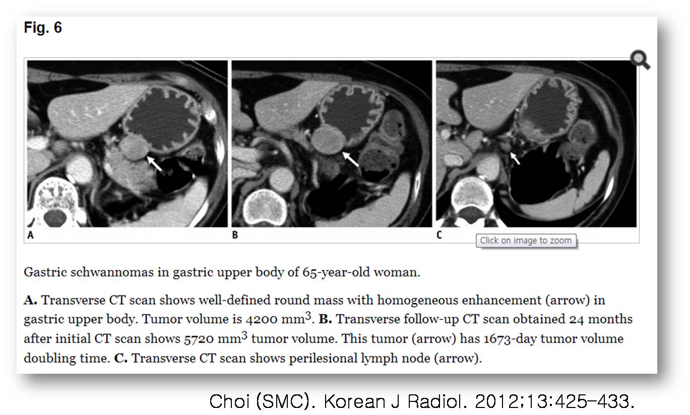
CT에서 schwannoma와 GIST를 감별하기는 쉽지 않습니다. 최근 삼성서울병원 영상의학과에서 의미있는 분석을 발표하였습니다 (Choi. Korean J Radiol. 2012;13:425-433). 신경초종은 GIST에 비하여 homogeneous enhancement, exophytic or mixed growth pattern, perilesional LN가 특징이었고 tumor volume doubling time은 GIST에서 377일, 신경초종은 1685일이었습니다.
병소 주변에 lymph node가 커진 경우가 많은 것이 Schwannoma의 특징입니다. 따라서 benign인데도 수술로 연결되는 경우가 많습니다. 림프절이 커져있으니 "혹시나?" 하는 생각이 들기 마련입니다.
Conclusion of the study: Although small gastric schwannomas (GSs) and GISTs show similar imaging findings, GSs more frequently show an exophytic or mixed growth pattern, homogeneous enhancement pattern, perilesional LNs and grow slower than GISTs.
Schwannoma인데 림프절이 뚜렸이 커졌던 증례를 소개합니다.
![]() 7. Esophageal schwannoma. 식도 신경초종
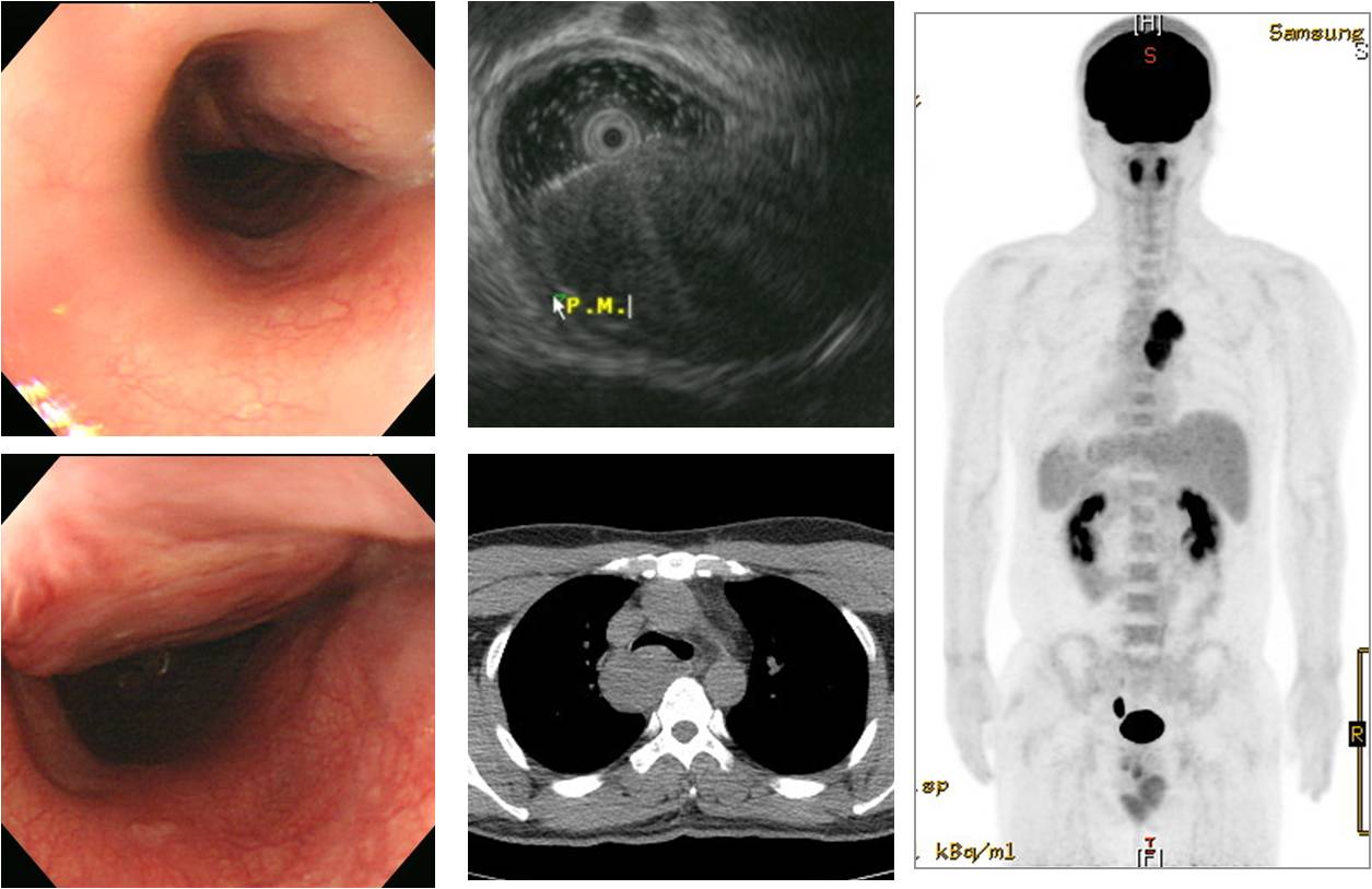
7. Esophageal schwannoma. 식도 신경초종
38대 남자로 2주전부터 연하곤란, 이물감으로 내시경을 시행하여 식도 종괴가 발견되었고 chest X-ray에서 mediastinal mass로 관찰되었습니다. VATS로 mass enucleation을 시행하였고 조직검사에서 Schwannoma, 5 x 3.5 cm, mitosis: up to 2/10 HPF, no necrosis의 소견이었습니다. 참고문헌을 인용합니다.
식도의 신경초종은 미주신경에서 발생한 경우와, 식도점막하의 신경총에서 발생한 경우로 분류될 수 있으며 전자의 경우, 미주신경에서 발생하여 식도벽으로 확장되는 양상을 보이며, 후자의 경우는 대개 식도점막하의 신경총에서 유래하여, 식도 근육층 내의 종양으로 나타난다. 식도에 발생하는 신경초종은 식도 조영술에서 부드러운 폴립같은 충만 결손을 보이며 식도 내시경에서는 돌출형 점막하 종양으로 보인다. 그러나 이것은 다른 식도 점막하 종양과 구별을 할 수 있는 특별한 소견이 아니므로 수술 전에는 식도 평활근종이나 낭종, 또는 지방종 등으로 진단되는 경우가 대부분이다. 진단에 있어서 종양의 형태를 쉽고 정확하게 측정할 수 있는 방법은 내시경 초음파이며, 흉부 전산화 단층촬영을 함께 이용하면 수술에 대한 정보를 더 얻을 수 있다. 일반적으로 방사선학적소견에 의해 낭종이나 지방종과의 구분은 쉽게 이루어질수 있다. 신경초종은 내시경적 초음파상 경계가 분명하고 저에코소견을 보이며, 고유 근육층에서 발생하여 점막 하에 위치하고, 흉부 전산화 단층 촬영상 연부조직종양의 형태로 보이며, 조영증강이 잘되지 않는다. (대흉외지 2005;38:589-593)
![]() 8. Colonic schwannoma. 대장 신경초종
8. Colonic schwannoma. 대장 신경초종
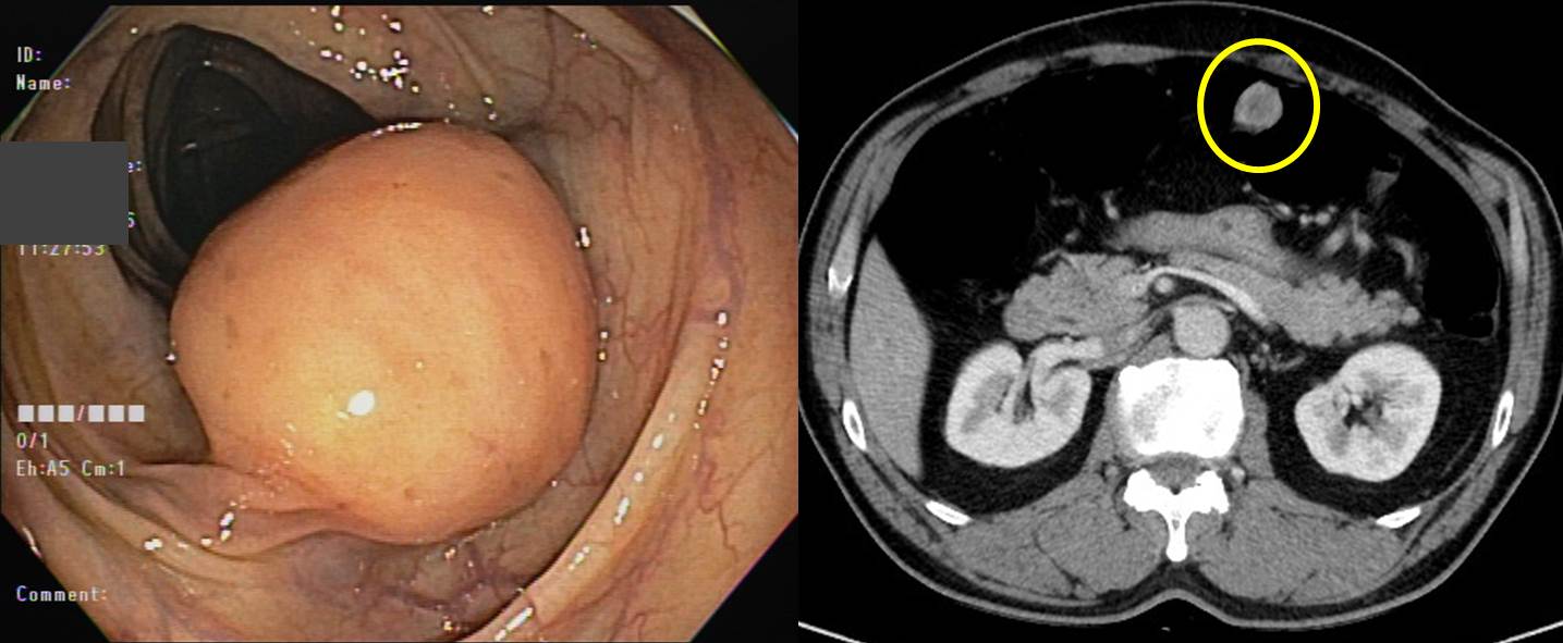
Hematochezia
검진 내시경 발견 RS junction SMT로 low anterior resection 시행함.
Schwannoma, plexiform type
2.3x2cm
12 reactive regional lymph nodes
![]() [References]
[References]
© 일원내시경교실 바른내시경연구소 이준행. EndoTODAY Endscopy Learning Center. Lee Jun Haeng.