EndoTODAY 내시경 교실
Beginner | ESA | Schedule | OPD
Seminars | Atlas | Recent | Links
![]() [Gastric diverticulum. 위(胃)게실] - 終
[Gastric diverticulum. 위(胃)게실] - 終
2020-4-25. 순천만내시경세미나 동영상 강의
![]() 1. Introduction to gastric diverticulum
1. Introduction to gastric diverticulum
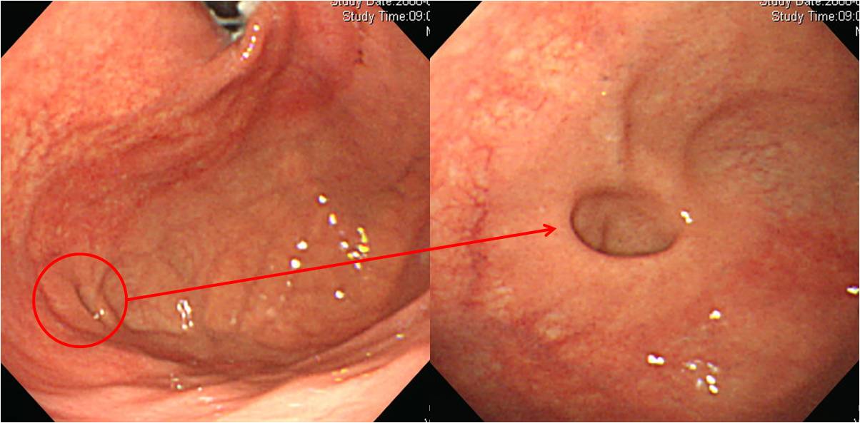
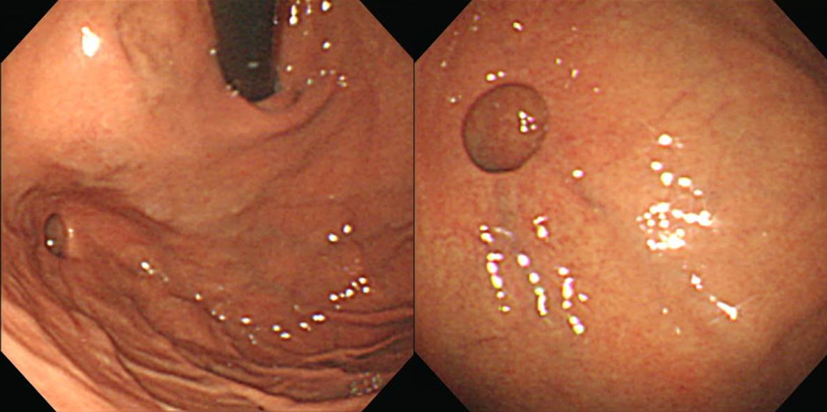
위 게실(gastric diverticulum)은 십이지장 게실이나 식도 게실에 비하여 드물게 발견됩니다. 대부분 무증상이므로 치료가 필요하지 않습니다. 드물게 출혈이 발생한 예가 보고되어 있습니다. 위암이 발생한 증례도 보았습니다 (Endoscopy 1998, Am J Gastroenterol 1987). 놀랄 일은 아닙니다. 게실 점막은 위점막이니 위암도 발생할 수 있겠지요. 큰 위게실이 증상을 일으켰다고 수술한 예도 있었습니다 JSLS 2006). 이 환자의 주증상에는 구취도 있었는데요... 사실 별로 믿어지지는 않습니다.
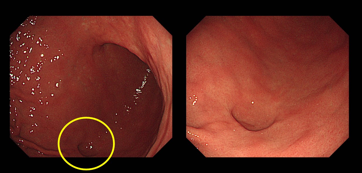
위의 큰 사진들은 위저부 게실의 내시경과 CT 소견입니다 (게실 때문에 찍은 CT는 아닙니다). 3장의 작은 사진들은 각기 다른 환자에서 발견된 게실이었습니다. 며칠 전에 말씀 드린 것 같지만, 게실은 한자로 憩室이라고 씁니다. '휴게실'의 '게실'입니다. 쉴 게, 방 실. 쉬는 방이라는 뜻입니다.
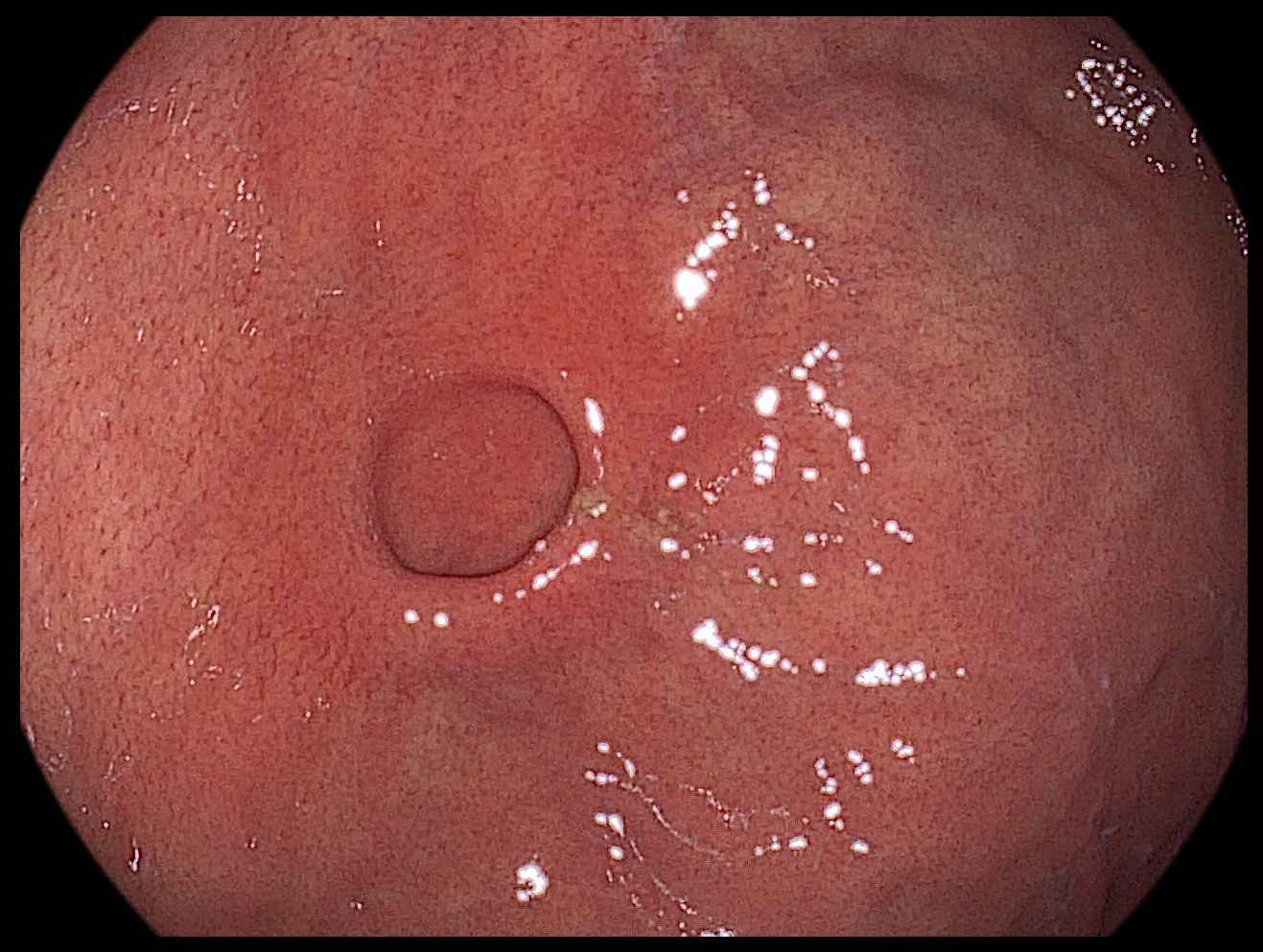
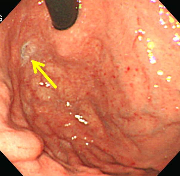
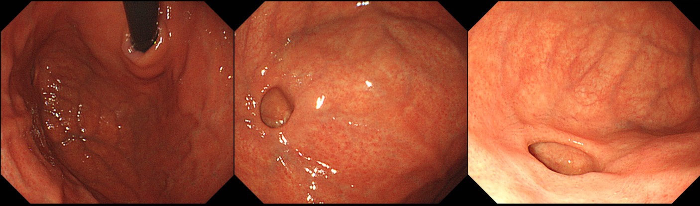
아직까지 저는 symptomatic gastric diverticulum 환자를 만난 적이 없습니다. 신경과 신장 합병증을 동반한 당뇨병을 가진 환자에서 비특이적 위장 증상으로 시행한 내시경 검사에서 작은 게실을 본 것이 전부입니다. 작고 깊지 않아서 증상과 관련이 있다고 생각되지는 않았습니다. (아래 증례)
F/75 (2017)
단기 PPI 사용 후 호전된 모습
위 게실은 주로 fundus에 많지만 드물게 다른 부위에서도 발견될 수 있습니다.
![]() 2. Pathophysiology of gastric diverticulum
2. Pathophysiology of gastric diverticulum
참고문헌을 옮깁니다. 게실이 왜 특정 위치에 잘 발생하는지에 대한 좋은 설명입니다.
위 게실은 소화기계 게실 질환들 중에서 가장 드문 형태로 검사 방법에 따라서 그 발생 빈도가 다양하게 보고되고 있는데, 상부 위장관 조영술에서 0.04%, 부검 연구에서는 0.02%, 상부위장관 내시경검사에서는 0.01∼0.11%에서 나타난다. 위 게실의 75% 이상은 위의 상부 후벽에서 발생하며, 이 위치는 식도-위 접합부의 약 2∼3 cm 하방에, 위 소만에서는 약 3 cm 떨어진 지점에 해당되며 해부학적으로 위 분문부의 종근층 섬유가 분리되어 있고 윤근층만이 점막을 덮고 있어서 게실 발생에 취약한 부위로 알려져 있다. 이 부위에서 발견되는 위 게실은 선천성이고, 진성 게실(true diverticulum)로서 위벽의 모든 층으로 구성되며 약 10%에서는 게실 내에 이소성 췌장 조직이나 위선 조직을 함유하는 것으로 알려져 있다. (김준성 등. 대한소화기내시경학회지 2007;34:99-102)
영문으로 된 리뷰(World J Emerg Surg 2012)에 실린 pathophysiology 설명입니다. Embryology로 설명하고 있습니다.
Seventy-five percent of true gastric diverticula were located in the posterior wall of the fundus of the stomach, 2 cm below the oesophagastric junction and 3 cm from the lesser curve. False diverticula were either traction or pulsion and associated with inflammation, other diseases, or both.
In the period between the 20th and 50th day of gestation, the stomach is transformed from a fusiform swelling of the foregut into its adult form. At this time, there is a 90° rotation of the stomach, which carries with it the duodenum, the pancreas, and the dorsal mesentery. The posterior body wall and dorsal mesentery then fuse encapsulating the pancreas within the retroperitoneum and establishing its adult form.
A diverticulum of the posterior wall of the gastric fundus hypothetically could herniate through an area of dorsal mesentery before its fusion with the left posterior body wall. Initially, the diverticulum would lie superior to the pancreas. With further extension, the diverticulum could project posterior to the pancreas.
Acquired gastric diverticula in contrast are pseudodiverticula, less common and typically located in the antrum. They usually present with a background history of other gastrointestinal pathology, such as peptic ulcer disease, malignancy, pancreatitis, or gastric outlet obstruction.
![]() 3. 위내시경 검사의 질지표로서 위게실
3. 위내시경 검사의 질지표로서 위게실
참고문헌을 찾을 수 없습니다만, 위게실의 발견율을 위내시경 검사의 질지표로 사용할 수 있다는 논문을 본 적이 있습니다. 위게실 자체가 무슨 중요한 의미가 있는 것은 아니지만 위게실을 잘 발견할 정도로 구석까지 정성껏 살폈다면 훌륭한 검사라는 의미일 것입니다.
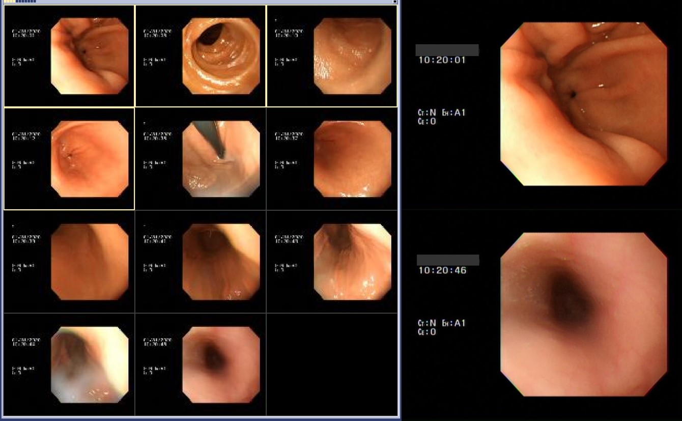
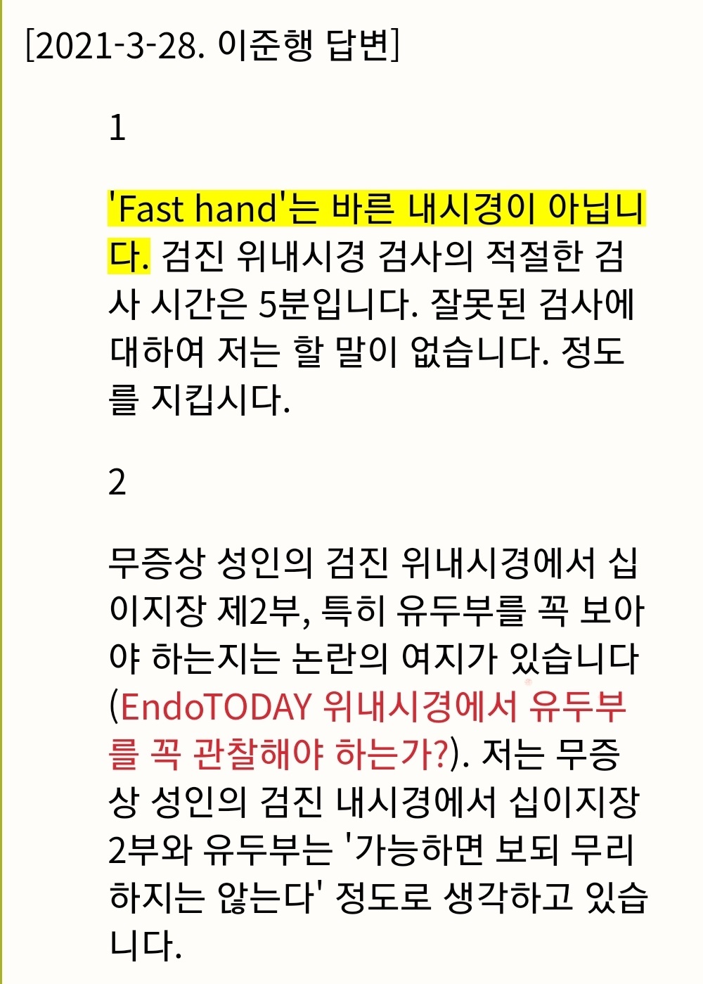
50대 남성입니다. 위게실을 발견하였습니다.
그런데 환자가 6개월 전 내시경 검사를 받았다고 하더군요. 그래서 과거 사진을 살펴보았습니다.
전정부가 첫 사진이라 정확히 검사 시간을 측정할 수 없었지만, 첫 사진은 10시 20분 1초이고 마지막 사진은 10시 20분 46초였습니다. 대부분 사진이 흔들리거나 흐린 상태였는데 contrast enhancement는 A1으로 되어 있었습니다. 너무하였다는 생각입니다.
빠른 내시경보다 바른 내시경을 합시다. 상부내시경 검사의 최소 검사 시간은 5분입니다.
![]() [Cases]
[Cases]
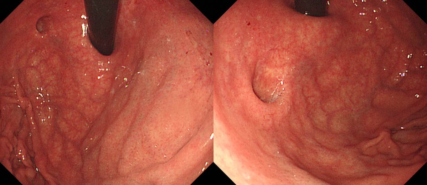
여러번 내시경하신 환자에서 우연히 gastric diverticlum을 발견하였습니다.
새로 생겼나 싶어서 과거 사진을 열어보니 과거부터 있었던 것 같습니다. 내시경을 할 때마다 "더 겸손해지자"고 반성합니다.

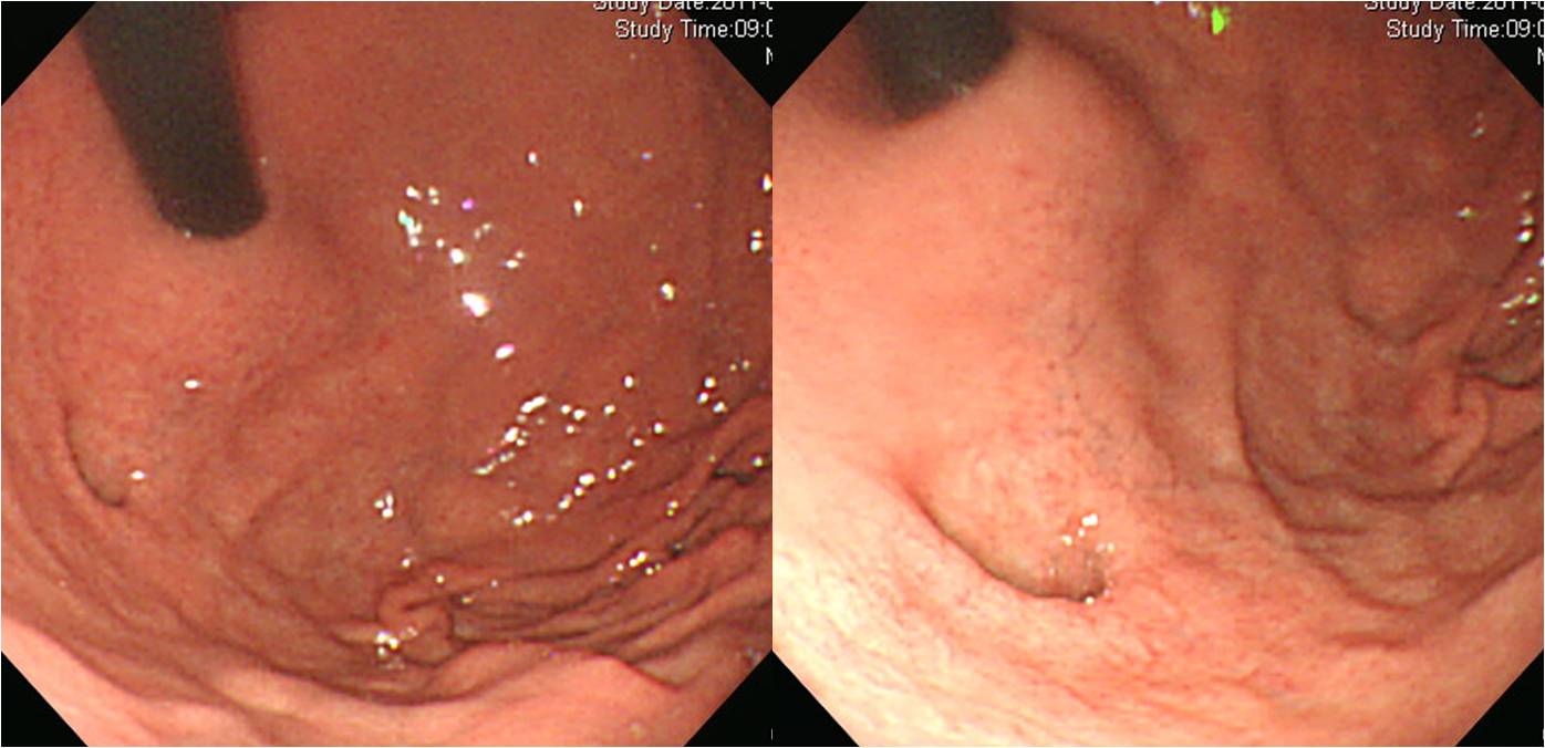
금식하고 내시경을 하였는데 다른 부위는 음식물이 없었으나 fundus에 약간의 음식물이 있었습니다. 게실에 음식이 고여있다가 뒤늦게 나온 것으로 판단되었습니다.
![]() [FAQ]
[FAQ]
[2021-3-30] fast hand?
얼마 전 어떤 질문을 받고 어이가 없어서 5분 검사시간을 강조하는 답변을 했습니다.
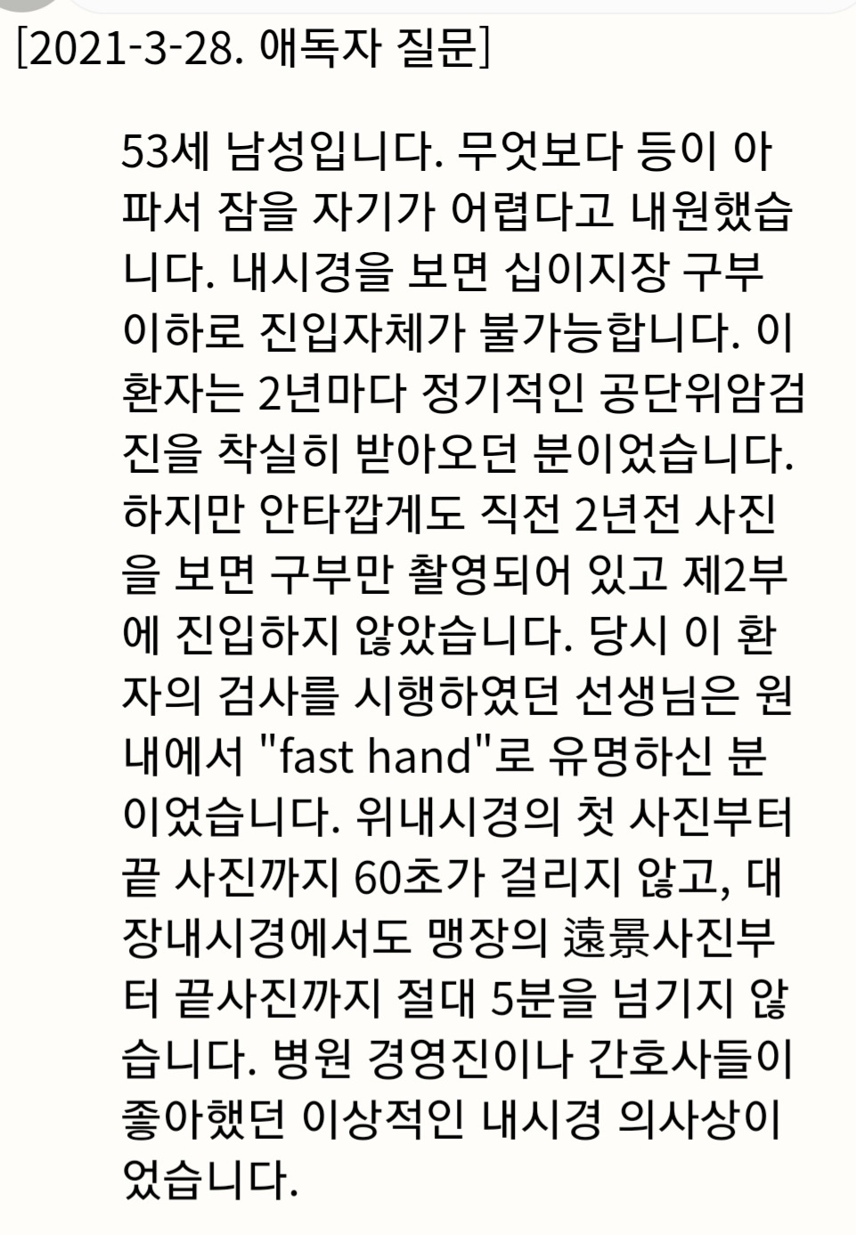
배울 때부터 제대로 배우고, 바르게 내시경 할 수 있는 제대로 된 환경을 만들고, 제대로 하고 있는지 monitoring 하는 것... 이 모든 것이 필요합니다. 안타깝습니다. 저수가 환경이 지겹습니다.
![]() [References]
[References]
1) 식도 게실: Zenker diverticulum, Killian-Jamieson 게실, 중부식도 게실, 하부식도 게실
2) 위 게실
3) 십이지장 게실
4) Meckel 게실
5) 대장 게실과 게실염
© 일원내시경교실 바른내시경연구소 이준행. EndoTODAY Endoscopy Learning Center. Lee Jun Haeng.