 EndoTODAY 내시경 교실
Beginner | ESA | Schedule | OPD
Seminars | Atlas | Recent | Links
 [위암 84]
001 | 101 | 201 | 301 | 401 | 501 | 601 | 701 | 801 | 901 | 1000
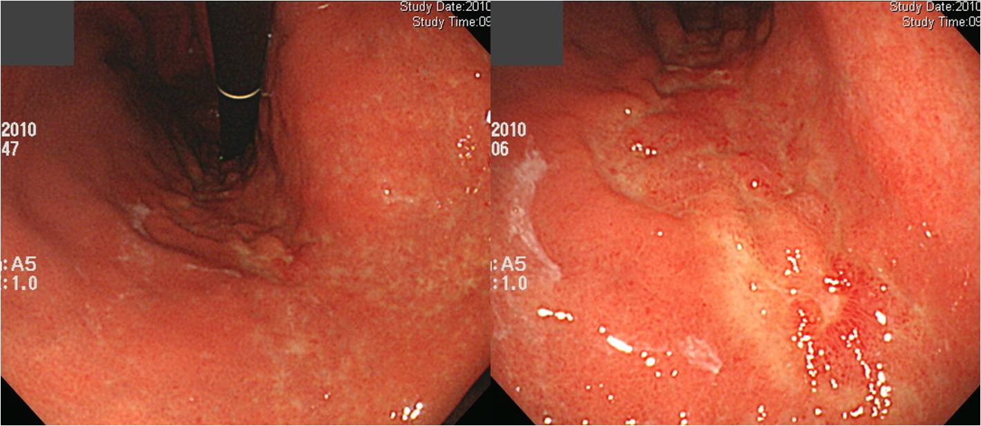
타병원에서 signet cell carcinoma로 진단받고 의뢰된 환자입니다. 이런 경우에 AGC Borrmann type을 어떻게 기술해야 하는지 궁금합니다. 또한 서술은 어떻게 해야 할지요?
아래로
아래로
아래로
아래로
아래로
 “타병원에서 signet cell carcinoma로 진단받고 의뢰된 환자입니다. 이런 경우에 AGC Borrmann type을 어떻게 기술해야 하는지 궁금합니다. 또한 서술은 어떻게 해야 할지요.”라는 질문을 받았습니다.
일견 5-6cm 정도의 depressed lesion이면서 base는 uneven하고 hyperemic한 부위와 pale discoloration을 보이는 부위가 mosaic pattern으로 관찰됩니다. 병소와 정상부위의 경계(edge)는 약간의 undulation을 보이지만 비교적 discrete합니다. 병소의 distal 부위에서는 명확하지 않으나 병소의 proximal 부위에는 abrupt cutting을 보이는 주름들이 모이는 양상입니다.
병소의 크기 때문에 AGC로 생각하신 것은 충분히 이해할 수 있지만 그래도 definite한 mass의 양상은 아닙니다. 작지 않은 병소이지만 그래도 superficial한 lesion이므로 EGC IIc로 주는 것이 좋겠습니다. 물론 proper muscle invasion이 있는 AGC Borrmann type unclassified 혹은 Borrmann type V라는 병리 리포트를 받게 되는 수가 있습니다. 그래도 내시경 의사는 EGC를 first impression으로 주는 것이 타당할 듯 싶습니다. Superficially spreading EGC 혹은 laterally spreading EGC로 부르기도 합니다.
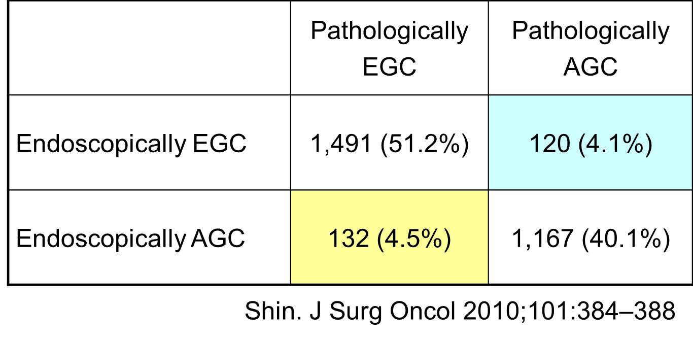
내시경 육안소견을 통하여 AGC와 EGC를 구분하는 것은 항상 틀릴 수 있는 일입니다. 일관성있는 태도를 견지하면서 약 90%의 정확도를 보이는 것이 목표가 아닐까 생각합니다. 아무리 주의해도 EGC와 AGC를 나누는 정확도는 80-90%전후입니다. 물론 아주 definite한 EGC가 있고 반박할 수 없는 AGC도 있습니다. 그러나 많은 경우 우리의 예상이 틀립니다. 그다지 놀랄 일은 아닙니다. 아래는 삼성서울병원의 결과 (Shin 2010)입니다.
[Related cases (EGC-like AGC)]
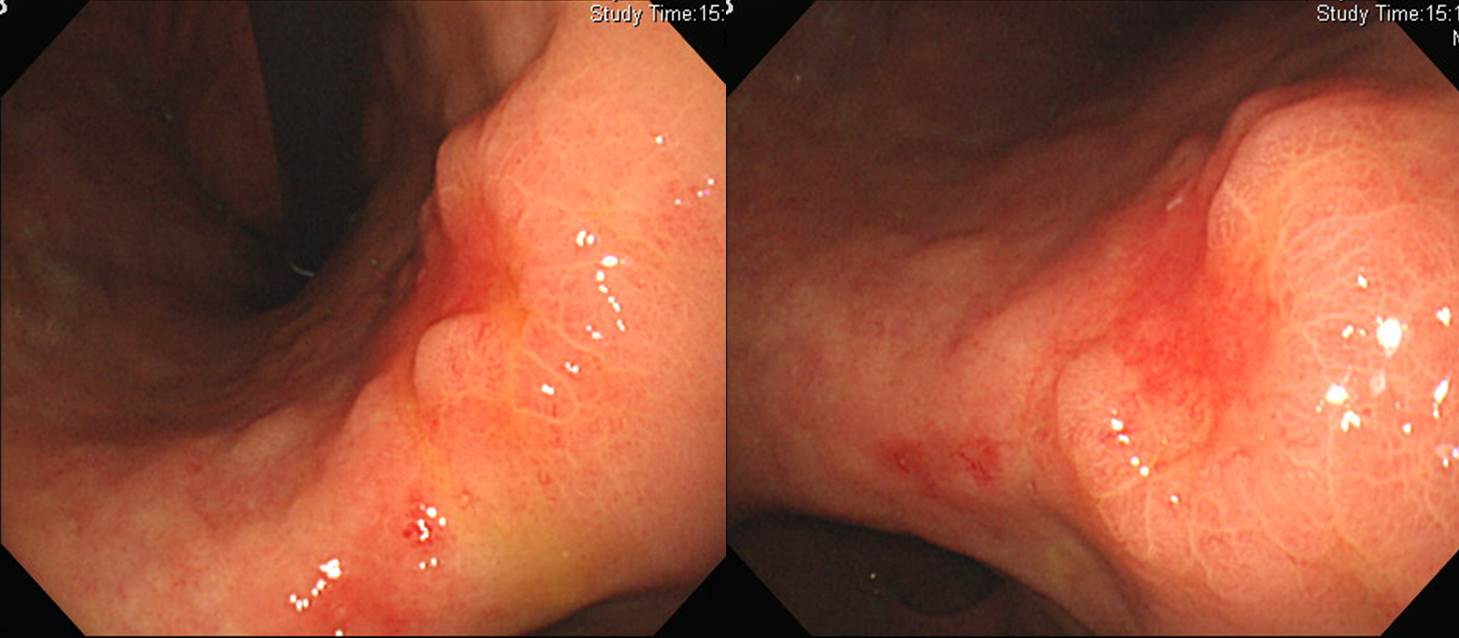
1. Location : middle third, center at body and posterior wall
2. Gross type : mimicking EGC type IIc
3. Histologic type : tubular adenocarcinoma, poorly differentiated
4. Histologic type by Lauren : diffuse
5. Size : 5x4x0.5 cm
6. Depth of invasion : extension to subserosa
7. Resection margin: free from carcinoma: safety margin
8. Lymph node metastasis : metastasis to 2 out of 35 regional lymph nodes (pN1)
9. Lymphatic invasion : present
10. Venous invasion : not identified
11. Perineural invasion : not identified
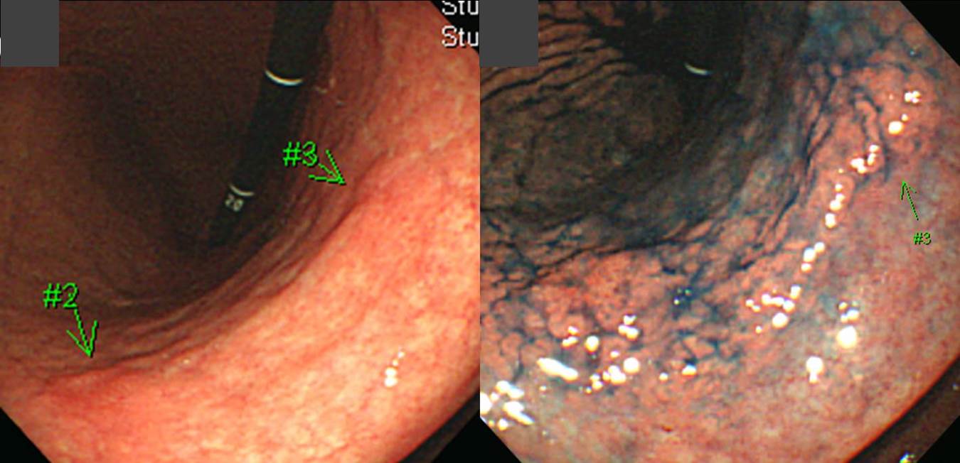
1. Location : middle third, center at body and lesser curvature
2. Gross type : Borrmann type (mimicking EGC type IIc+III)
3. Histologic type : signet-ring cell carcinoma
4. Histologic type by Lauren : diffuse
5. Size : 9.5x5.3x0.4 cm
6. Depth of invasion : extension to proper muscle (pT2a)
7. Resection margin: free from carcinoma: safety margin
8. Lymph node metastasis : metastasis to 9 out of 62 regional lymph nodes
9. Lymphatic invasion : present
10.Venous invasion : not identified
11.Perineural invasion : not identified
12.Associated findings : ulceration(ul IV)
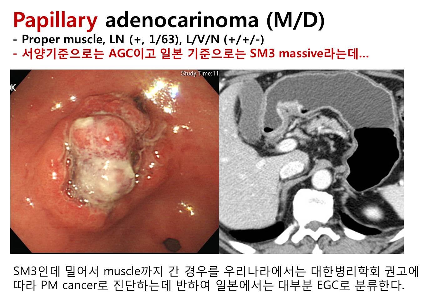
조기위암과 진행성위암의 구분이 항상 명확한 것은 아닙니다. 우리나라보다 일본의 암진단 기준이 더 낮은 것은 유명한 일입니다. 조금만 이상해도 일본에서는 암으로 진단붙여버리니... 참 속편한 사람들입니다. 우리나라의 high grade dysplasia가 일본에 가면 대부분 암으로 진단되고 있다고 여겨집니다. 그런데 반대의 상황도 있습니다. 우리나라에서 AGC로 분류하는데 일본에서는 EGC로 분류하는 그런 상황도 있는 것입니다. 두 나라의 병리학자들은 확실히 다릅니다.
2014. 6. 23. 이준행