 EndoTODAY 내시경 교실
Beginner | ESA | Schedule | OPD
Seminars | Atlas | Recent | Links
 [위암 296]
001 | 101 | 201 | 301 | 401 | 501 | 601 | 701 | 801 | 901 | 1000
과거 위암으로 subtotal gastrectomy 받으신 분께서 cardia polyp의 조직검사에서 adenoma가 나왔다고 의뢰되었습니다. 그러나 모양은 hyperplastic polyp과 구분이 어려운 용종입니다. Size가 다소 크고 표면도 불규칙하고 friability도 있어서 암의 가능성이 높다고 생각되었습니다. Adenoma가 이런 모습인 경우는 거의 없습니다.
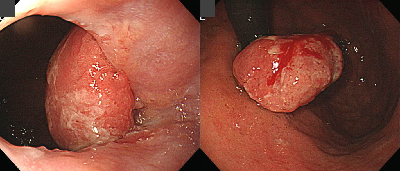
조직검사 재검에서 암이 나와 ESD가 시행되었습니다. 5년 이상 되었는데 아직 아무 문제는 없습니다.
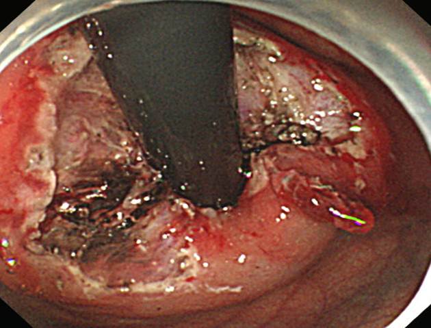
Stomach, endoscopic submucosal dissection:
Early gastric carcinoma
1. Location : cardia
2. Gross type : EGC type I
3. Histologic type : tubular adenocarcinoma, well differentiated
4. Histologic type by Lauren : intestinal
5. Size : 3.5x2.8x1 cm
6. Depth of invasion : invades mucosa (muscularis mucosa) (pT1a)
7. Resection margin: free from carcinoma, safety margin: distal 0.2 cm, proximal 0.1 cm, anterior 0.2 cm, posterior 0.1 cm
8. Lymphatic invasion : not identified
9. Venous invasion : not identified
10. Perineural invasion : not identified
11. Microscopic ulcer : absent
12. Histologic heterogeneity: absent
Hyperplastic polyp 양상인데 조직검사에서 adenoma가 나오면 선종일 것으로 추정하는 것 보다는 "조직검사는 neoplastic lesion인 것으로 나왔으니 암일 확률이 높겠군..." 정도로 생각하시는 것이 좋겠습니다.
© 일원내시경교실 바른내시경연구소 이준행. EndoTODAY Endoscopy Learning Center. Lee Jun Haeng.