Previous | Home | EndoTODAY | List | Next
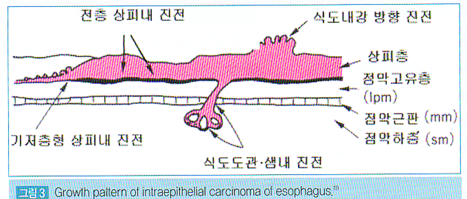
![]() [Growth pattern of intraepithelial carcinoma of esophagus]
[Growth pattern of intraepithelial carcinoma of esophagus]
간혹 식도 상피암이 gland를 따라서 점막하층까지 spread 되는 경우가 있다고 합니다.
매우 드문 일이라고 생각합니다. ESD를 시행한 환자에서 딱 한번 경험한 적이 있을 뿐입니다.
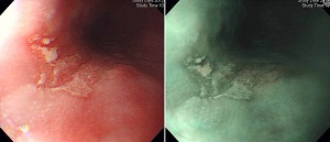
Squamous cell carcinoma, in-situ :
1) size: up to 12 mm
2) marked glandular extension
3) no involvement of margin (see note)
Note: In some section, tumor cell nest of glandular extension (not true invasion) is very close to deep margins.
![]() [2014-12- 26 추가] 2015년 1월 Gastrointestinal Endoscopy에 식도암 소작술의 위험성을 경고하는 논문이 실렸습니다. 미국과 일본의 의사들이 함께 쓴 논문입니다 (link). 소작술 적응증인 병소를 내시경 절제하면 생각보다 깊거나 림프선 침윤을 보이는 경우가 있으니 주의하라는 이야기였습니다. 동의합니다. 내시경 절제술이 소작술보다 질병의 치료면에서는 분명 좋습니다. 단지 위험할 뿐이지요.
[2014-12- 26 추가] 2015년 1월 Gastrointestinal Endoscopy에 식도암 소작술의 위험성을 경고하는 논문이 실렸습니다. 미국과 일본의 의사들이 함께 쓴 논문입니다 (link). 소작술 적응증인 병소를 내시경 절제하면 생각보다 깊거나 림프선 침윤을 보이는 경우가 있으니 주의하라는 이야기였습니다. 동의합니다. 내시경 절제술이 소작술보다 질병의 치료면에서는 분명 좋습니다. 단지 위험할 뿐이지요.
이 논문에 점막의 표재성 식도암으로 판단되었는데 ESD 후 marked glandular extension을 보인 경우가 소개되었습니다. 병리사진이 좋아서 옮깁니다.
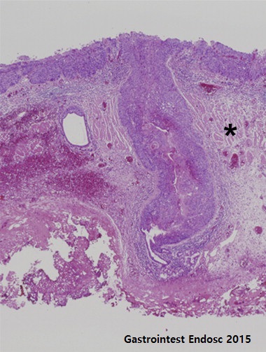
Ductal extension in an endoscopic submucosal dissection specimen. Representative example of a T1m2 lesion showing massive ductal extension. The superficial component of the lesion shows an undulating, expansive pattern of growth, consistent with T1m2. The asterisk indicates the muscularis mucosae. The lesion extends via the ductal system to the level of the submucosal gland (link)
필자도 이와 비슷한 경우 경험하여 2013년 1월 14일 EndoTODAY에 소개한 바 있어 아래에 옮깁니다.
[Home]
 크게
크게