EndoTODAY 내시경 교실
Beginner | ESA | Schedule | OPD
Seminars | Atlas | Recent | Links
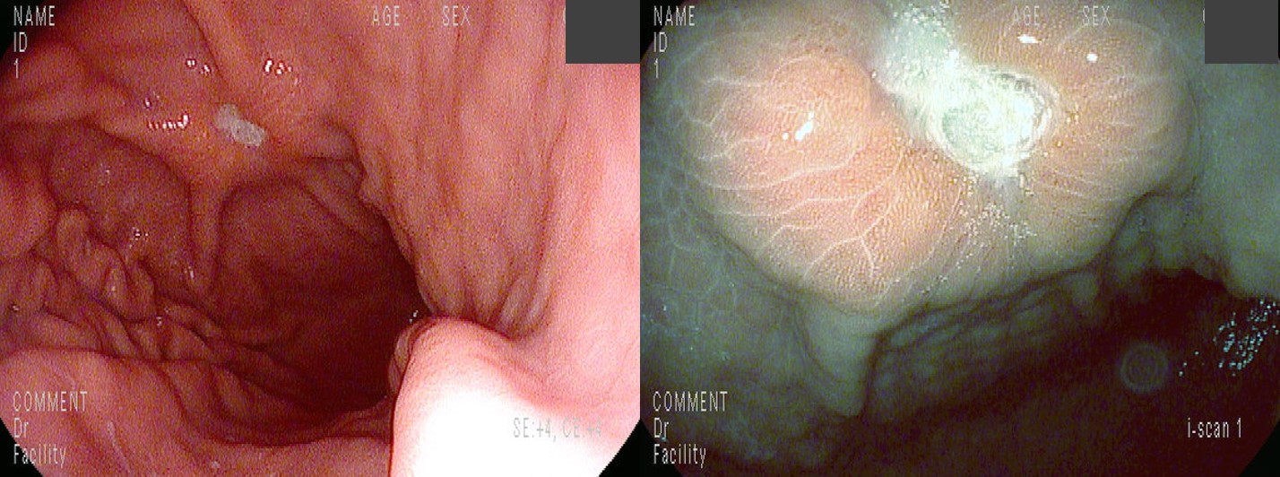
 [Gastric cancer 944. Focal wall-thickening mass with central ulceration and converging folds] - 終
001 | 101 | 201 | 301 | 401 | 501 | 601 | 701 | 801 | 901 | 1000
P/D with signet ring cell component라는 조직검사 결과와 함께 의뢰된 분입니다.
의뢰 후 재검을 하였고 거의 동일한 소견이 보였습니다.
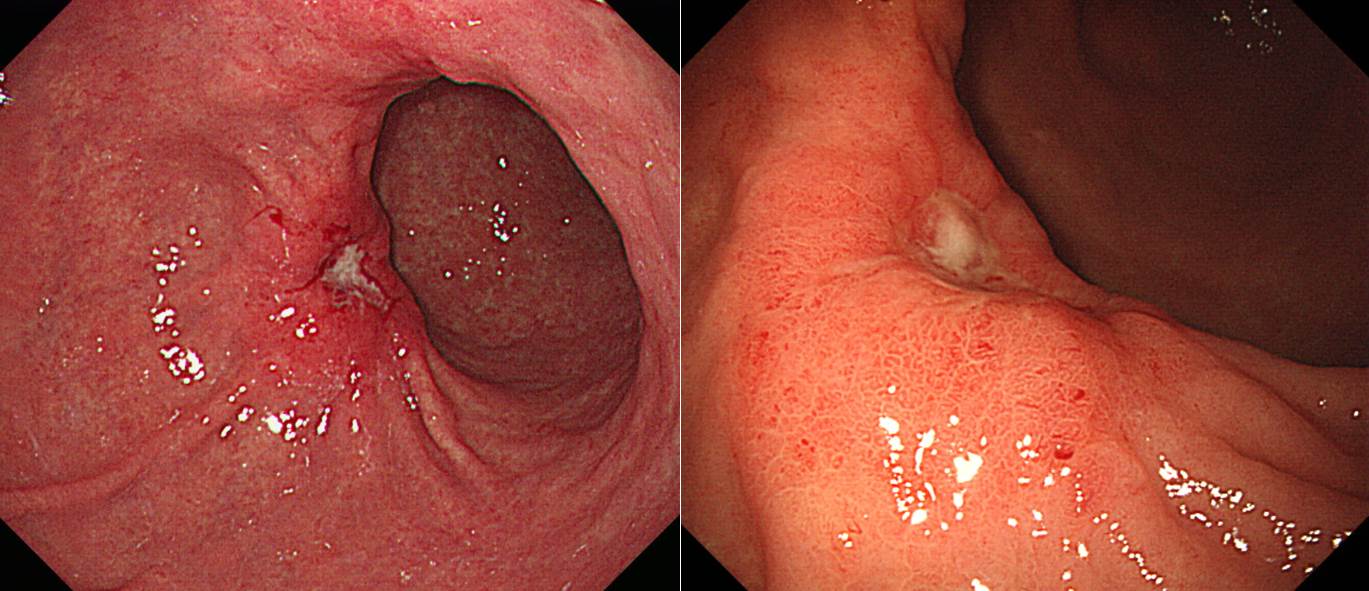
내시경 소견은 focal wall-thickening mass with central ulceration and converging folds로 요약할 수 있습니다.
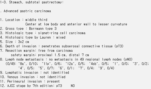
수술을 시행하였습니다.
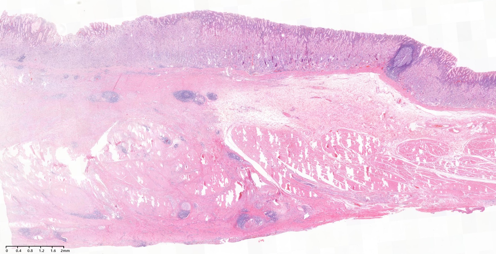
Focal wall thickening을 보인 AGC였습니다. Subserosal penetration이 있었으나 serosal invasion은 없었습니다. 림프절 전이는 없었습니다.
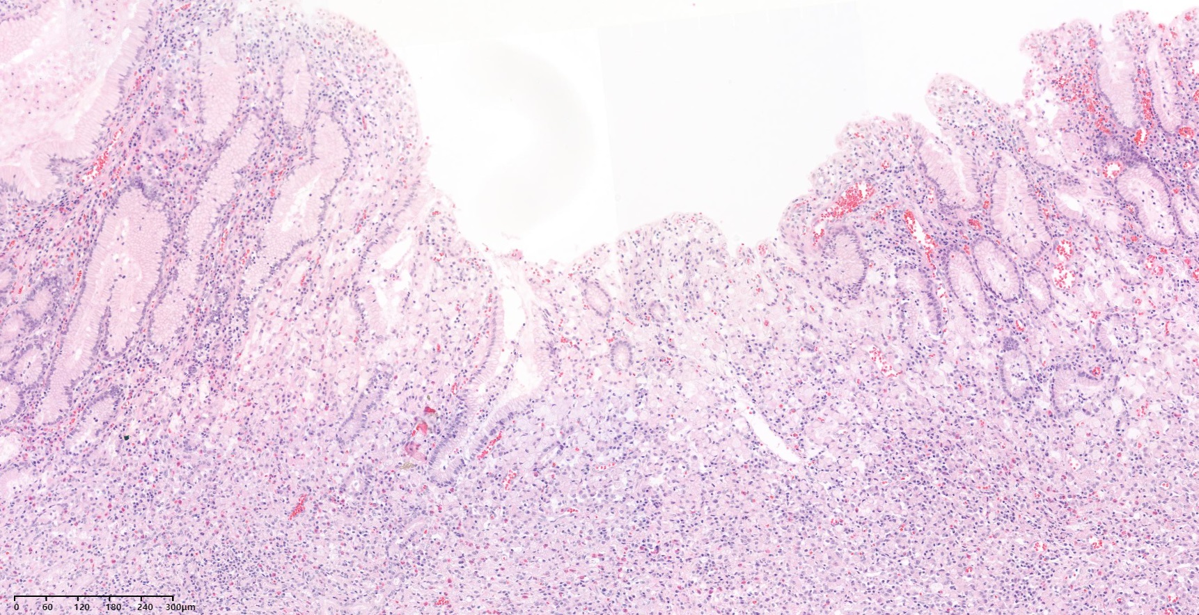
일부 점막 함몰부에서는 표면에 암세포가 노출되었습니다.
다른 부위에서는 foveolar epithelium 바로 아래 mid-to lower lamina propria부터 암세포가 보였습니다. 내시경 소견에서 wall-thickening mass with central ulceration과 일치하는 병리소견입니다.
 [2024-11-9] 증례토의/퀴즈 중 한 fellow께서 아래와 같은 impression을 주었습니다.
"Lower body anterior wall에 3cm의 elevated mass with central ulceration이 관찰되며 주위 점막의 끌어담김이 관찰되어 SMT의 가능성은 낮아보이고 AGC라 하기엔 중심부 궤양의 모양이 비교적 깨끗하여 lymphoma 가능성이 높다고 생각합니다"
이준행 comment: 소견은 잘 기술하셨습니다. 그런데 impression이 틀렸군요. 주 소견이 mass이고 top central ulceration이 있고 fold가 ulcer까지가 아니고 mass의 경계까지 끌려오는 상황입니다. DLBCL와 MALT lymphoma와의 내시경 소견은 제법 다릅니다. DLBCL는 종양의 성장 속도가 빠르고 desmoplastic reaction = fibrosis가 거의 동반되지 않기 때문에 fold 변화를 일으키지 않습니다. 반면 MALT lymphoma는 악화와 호전을 반복하는 특징이 있어서 EGC나 BGU scar 처럼 fold 변화를 보이기도 합니다. 여하튼 이 증례는 mass의 periphery까지 다수의 fold가 끌려오고 있으므로 lymphoma의 가능성은 매우 매우 낮을 것으로 보입니다.
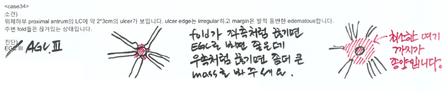
Description Exercise Quiz 5회의 34번 증례가 이 환자와 매우 비슷합니다. 정답률이 낮은 문제입니다.
소견: Mid-antrum AW에 4cm 정도의 mass가 있습니다. 병변의 중앙에는 ulcer가 있으며 margin은 hyperemic, edematous하고 edge는 spiculation 되어 있습니다. 병변 방항으로 fold가 여러개 끌려오는 것이 관찰됩니다.
진단: AGC, B-III
[이준행 comment]
병변의 주된 특징을 ulcer로 보고 EGC라 답한 분이 계셨습니다. 병변의 주된 특징을 ulcer로 보지 말고 mass로 보고, mass 중앙에 작고 깊은 ulceration이 있다고 보면 더 좋겠습니다. 즉, '궤양 1cm + 주변 부종'으로 보는 것도 좋지만 '4cm mass + 중앙 궤양'으로 보면 어떤가 합니다. 그리고 fold 끊긴 곳의 위치도 잘 보세요.
주(主)소견, 즉 major finding을 정확히 잡는 것은 매우 중요한 일입니다.
수술을 하였고 생각보다 깊었습니다.
Stomach, subtotal gastrectomy:
. Advanced gastric carcinoma
1. Location : middle third, Center at body and anterior wall
2. Gross type : Borrmann type 3
3. Histologic type : signet-ring cell carcinoma
4. Histologic type by Lauren : diffuse
5. Size : 6.5x4.5 cm
6. Depth of invasion : penetrates subserosal connective tissue (pT3)
7. Resection margin: free from carcinoma, safety margin: proximal 2.5 cm, distal 3.5 cm
8. Lymph node metastasis : metastasis to 4 out of 67 regional lymph nodes (pN2), (perinodal extension: present)
9. Lymphatic invasion : present
10. Venous invasion : not identified
11. Perineural invasion : present
12. Peritoneal cytology : negative
13. AJCC stage by 7th edition: pT3 N2
이 증례를 포함하여 유사한 증례를 동영상 강의로 설명합니다.

© 일원내시경교실 바른내시경연구소 이준행. EndoTODAY Endoscopy Learning Center. Lee Jun Haeng. (2024-11-7)