EndoTODAY 내시경 교실
Beginner | ESA | Schedule | OPD
Seminars | Atlas | Recent | Links
 [Gastric cancer 903. 넓은 EGC IIc]
001 | 101 | 201 | 301 | 401 | 501 | 601 | 701 | 801 | 901 | 1000
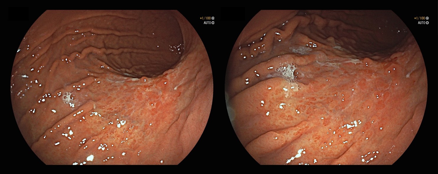
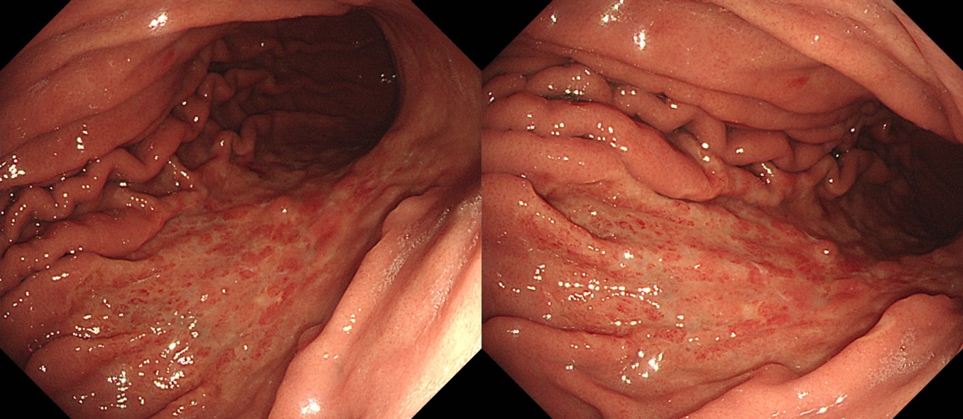
Fujifilm 내시경으로 시행된 건진 내시경에서 위암이 발견되어 의뢰되었습니다. 외부 조직검사 재판독은 tubular adenocarcinoma, poorly differentiated with signet ring cell feature였습니다. 침윤 깊이는 어떻게 예측하십니까? 내시경 분류는 무엇이라고 생각하십니까?
Olympus 내시경으로 재검을 하였고 동일 소견이었습니다.
수술을 시행하였습니다.
Stomach, radical total gastrectomy: Early gastric carcinoma
1. Location : middle third, Center at body and posterior wall
2. Gross type : EGC type IIc
3. Histologic type : tubular adenocarcinoma, poorly (poorly cohesive) differentiated
4. Histologic type by Lauren : mixed
5. Size : 7x6.5 cm
6. Depth of invasion : invades mucosa (muscularis mucosae) (pT1a)
7. Resection margin: free from carcinoma, safety margin: proximal 3.5 cm, distal 9 cm
8. Lymph node metastasis : no metastasis in 42 regional lymph nodes (pN0) (0/42: "3,5", 0/11; "4,6", 0/5; "1", 0/2; "11p", 0/2; "12a", 0/2; "4sb", 0/0; "5", 0/1; "6", 0/3; "7", 0/3; "8a", 0/2; "9", 0/7; "2", 0/3; "perigastric LN", 0/1)
9. Lymphatic invasion : not identified
10. Venous invasion : not identified
11. Perineural invasion : not identified
12. Peritoneal cytology : negative
13. AJCC stage by 8th edition: pT1a N0
. c-erbB-2 : Negative (1+)
. MLH1 : Intact
. Epstein-Barr virus: Negative
비교적 큰 병소였지만 flat depressed lesion이고 mass 소견이 없고 위벽이 잘 펴지는 양상이라 조기위암임은 짐작할 수 있었습니다. 크다고 깊은 것은 아닙니다. 그러나 이 정도 병소이고 미분화조직형인 경우는 국소적인 SM invasion인 경우가 많아서 점막하암일 것으로 추정하였습니다만, 결과는 muscularis mucosa까지만 침윤된 암이없습니다. 심달도 예측은 참으로 어렵습니다. 조기위암 분류는 IIc가 적당합니다. 전체적으로 flat depressed lesion이었지만 fold가 끊기면서 약간 내려 앉은 모양 아닙니까? 병리과에서도 IIc로 주었네요.
© 일원내시경교실 바른내시경연구소 이준행. EndoTODAY Endoscopy Learning Center. Lee Jun Haeng. (2021-9-22)