EndoTODAY 내시경 교실
Beginner | ESA | Schedule | OPD
Seminars | Atlas | Recent | Links
 [Gastric cancer 865. Focal non-atrophic area]
001 | 101 | 201 | 301 | 401 | 501 | 601 | 701 | 801 | 901 | 1000
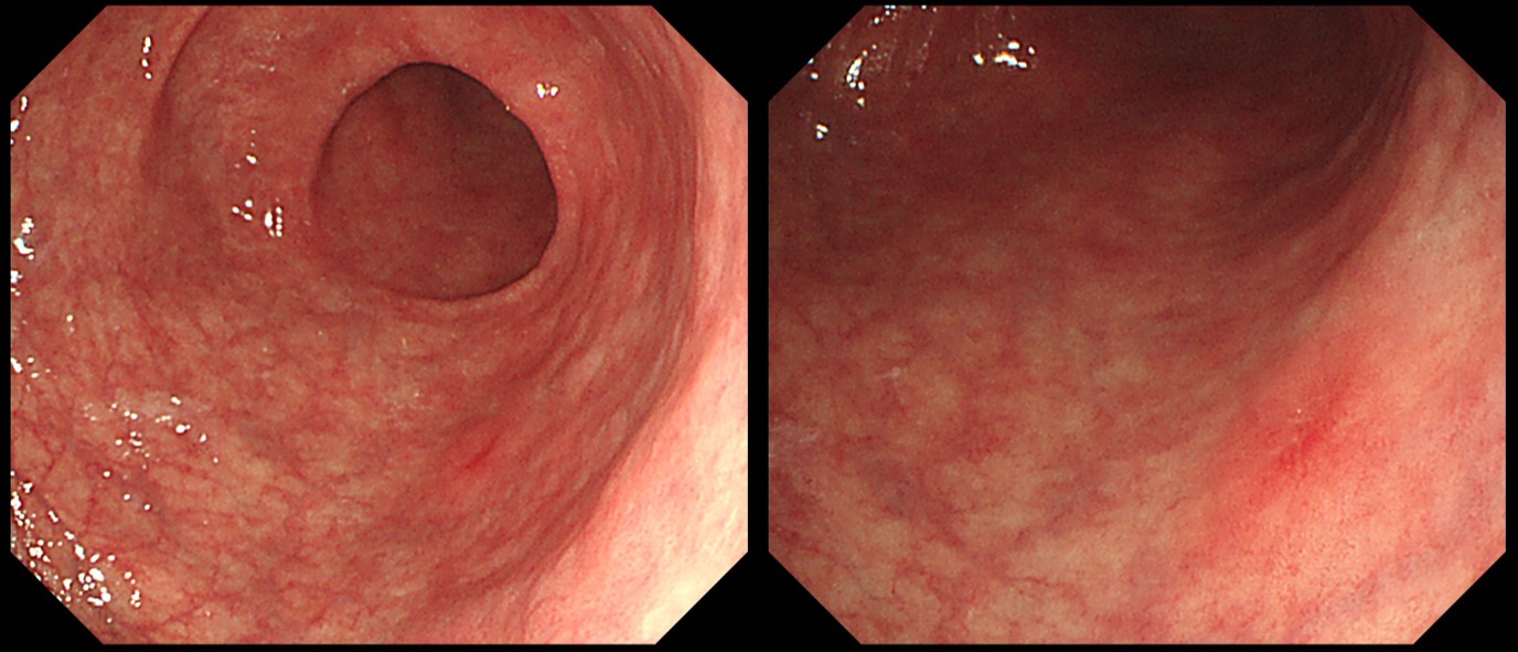
65 years old female was referred due to an EGC (biopsy: moderately differentiated adenocarcinoma). It was a small non-atrophic area in the GC side of the proximal antrum. In the close-up image, it was a small flat elevated lesion with slight depression in the center.
ESD was done as usual.
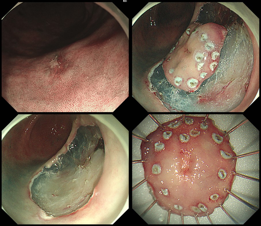
ESD: Early gastric carcinoma
1. Location : antrum, greater curvature
2. Gross type : EGC type IIc
3. Histologic type : tubular adenocarcinoma, moderately differentiated
4. Histologic type by Lauren : intestinal
5. Size of carcinoma : (1) longest diameter, 7 mm (2) vertical diameter, 6 mm
6. Depth of invasion : invades mucosa (muscularis mucosa) (pT1a)
7. Resection margin : free from carcinoma(N), safety margin : distal 10 mm, proximal 8 mm, anterior 10 mm, posterior 12 mm, deep 500 ㎛
8. Lymphatic invasion : not identified(N)
9. Venous invasion : not identified(N)
10. Perineural invasion : not identified(N)
11. Microscopic ulcer : absent
12. Histologic heterogeneity: absent
[우리말 註]
가까이 본다고 잘 보이는 것은 아닙니다. 멀리서 위축된 점막을 고루 관찰하며 non-atrophic area가 없는지 살피면 작은 위암을 발견할 수 있습니다.
© 일원내시경교실 바른내시경연구소 이준행. EndoTODAY Endoscopy Learning Center. Lee Jun Haeng. (2020-6-14)