EndoTODAY ł»˝Ă°ć ±ł˝Ç
Beginner | ESA | Schedule | OPD
Seminars | Atlas | Recent | Links
![]() [Gastric cancer 824. ESD for serrated adenocarcinoma of the stomach]
[Gastric cancer 824. ESD for serrated adenocarcinoma of the stomach]
001 | 101 | 201 | 301 | 401 | 501 | 601 | 701 | 801 | 901 | 1000
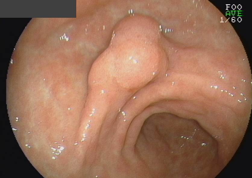
A patient was referred due to tubulovillous adenoma with high grade dysplasia of the antrum. The result of the outside pathology slide was "serrated adenocarcinoma, well differentiated." ESD was done.

ESD: Early gastric carcinoma
1. Location : antrum, lesser curvature
2. Gross type : EGC type IIa
3. Histologic type : serrated adenocarcinoma, well differentiated
4. Histologic type by Lauren : intestinal
5. Size of carcinoma : (1) longest diameter, 6 mm (2) vertical diameter, 4 mm
6. Depth of invasion : invades mucosa (lamina propria) (pT1a)
7. Resection margin : free from carcinoma(N), safety margin : distal 10 mm, proximal 6 mm, anterior 16 mm, posterior 16 mm, deep 800
8. Lymphatic invasion : not identified(N)
9. Venous invasion : not identified(N)
10. Perineural invasion : not identified(N)
11. Microscopic ulcer : absent
12. Histologic heterogeneity: absent
[ ]
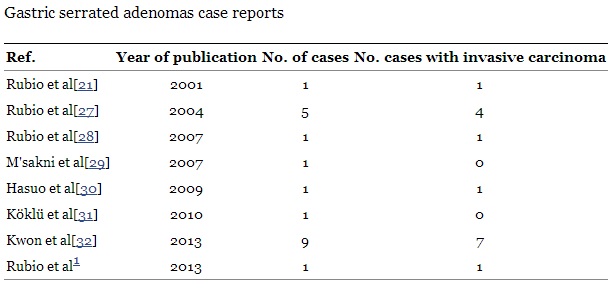
serrated adenoma ? serrated adenoma Ąď ?? . 2001 Sweden Carlos Rubio u 2013 20 ? ? . ? ? Kwon et al ŁÚ ? ? ?? .
World J Gastrointest Endosc. 2013 May 16;5(5):261-4
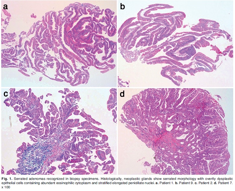
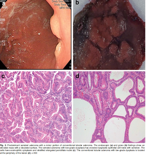
? ??? ? ? ? Kwon Ąę ? ? ? ? . "The gastric serrated adenoma has elongated fronds with lateral crenated, saw tooth-like notches as a result of scalloped epithelial indentations. The serrated crypts are lined by overtly dysplastic epithelial cells containing abundant eosinophilic cytoplasm and stratified elongated penicillate nuclei. These microscopic features mimic traditional serrated adenoma found in the colorectum." ? ? ? ? ? ? ? ?? . ?? ŻC ©Ł ? ? " ? ? " ? ? ? . (Histol Histopathol. 2013 Apr;28(4):453-62)
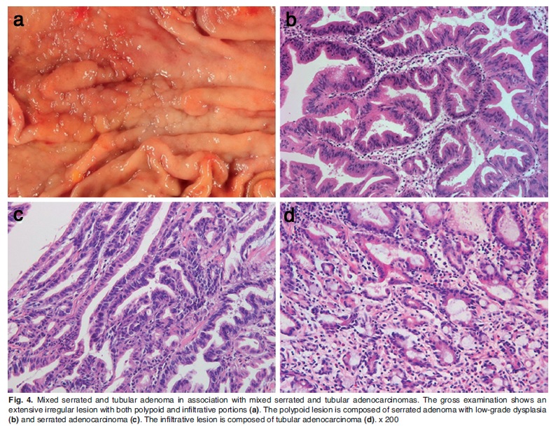
2013 o serrated adenocarcinoma ©Ł ? ?? .
2018 ESD ? ? ?? .
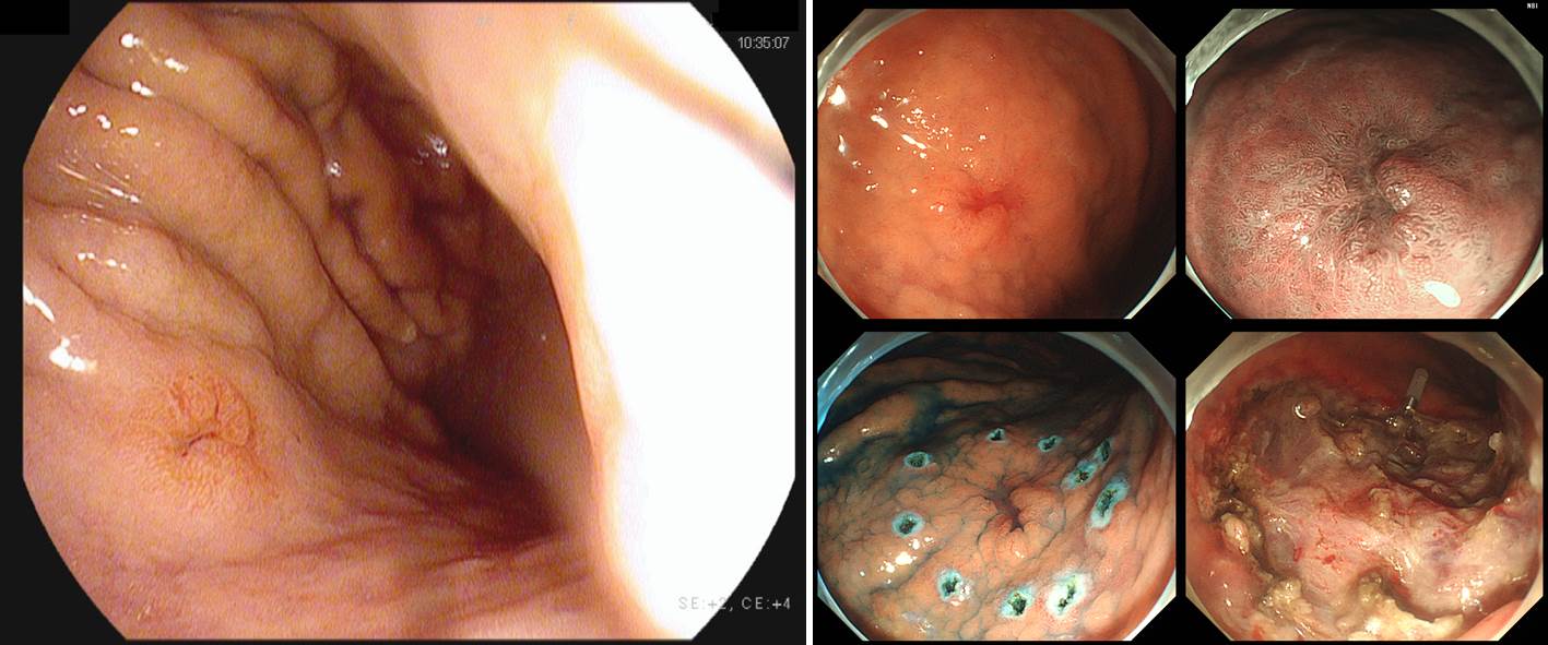
ESD : Early gastric carcinoma
1. Location : high body, posterior wall
2. Gross type : EGC type IIc
3. Histologic type : tubular adenocarcinoma, moderately differentiated with serrated feature
4. Histologic type by Lauren : intestinal
5. Size of carcinoma : (1) longest diameter, 6 mm (2) vertical diameter, 6 mm
6. Depth of invasion : invades submucosa, (depth of sm invasion : 1200 ) (pT1b)
7. Resection margin : involved deep resection margin by carcinoma with cauterized artifact negative other resection margins; safety margin : distal 3 mm, proximal 2 mm, anterior 16 mm, posterior 4 mm, deep 0
8. Lymphatic invasion : not identified(N)
9. Venous invasion : not identified(N)
10. Perineural invasion : not identified(N)
11. Pre-existing adenoma : none
12. Microscopic ulcer : absent
13. Histologic heterogeneity: absent
Becuase of the deep SM invasion and positive vertical margin, additional surgical treatment was recommended.
The meaning of the serrated feature in the gastric neoplasm is still unclear.
© ŔĎżřł»˝Ă°ć±ł˝Ç ąŮ¸Ął»˝Ă°ćż¬±¸ĽŇ ŔĚÁŘÇŕ. EndoTODAY Endoscopy Learning Center. Lee Jun Haeng. (2020-1-29)