EndoTODAY 내시경 교실
Beginner | ESA | Schedule | OPD
Seminars | Atlas | Recent | Links
 [Gastritis cystica. 낭성 위염] - 終
 1. Gastritis cystica profunda (GCP). 심재성 낭성 위염
선상피의 과형성 및 낭성 확장으로 위점막의 심부 및 점막하층에 낭종을 형성하는 질환입니다. 대개 위절제술을 시행 받은 환자에서 발생하나 드물게는 위 수술의 기왕력이 없는 경우에도 보고되고 있습니다. 병변은 점막하 종양, 용종, 간혹 거대 점막 주름 형태로 나타날 수 있으며 일반적으로 전암 병변이 아닌 것으로 받아들여지고 있으나, GCP의 악성화 경향이나 악성과의 관련성에 대해서는 논란이 있습니다.
gastritis cystica in the fundus. Inject and cut 방법으로 치료.
gastritis cystica in the fundus. Water immersion EMR 방법으로 치료 (위질환내시경아틀라스 7호에 실렸던 부산대 증례)
2. 암과 함께 발견된 gastritis cystica profunda (GCP)
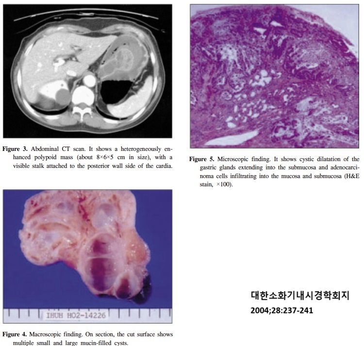
2004년 인하대에서는 GCP에서 기원한 위선암 1예를 보고한 바 있습니다 (대한소화기내시경학회지 2004).
대부분이 GCP이고 일부에서 암인 경우보다는 위암의 일부에서 GCP가 동반된 예가 더 많은 것 같습니다. 아래는 Deep SM layer까지 침윤된 위암입니다. 병리과 선생님께 문의하였고 다음과 같은 답변을 받았습니다. "정상 gland의 herniation, gastritis cystica profunda로 보면 될 것 같습니다. GCP는 다양한 gland, 양으로 구성될수 있고, 사진에서 주변에 lamina propria도 약간 있습니다. 책에는 보통 lamina propria가 둘러싸고 있다고 되어 있지만, gland만 herniation되는 경우도 있습니다. GCP는 normal mucosa뿐 아니라 이 증례처럼 종종 암조직에서 동반되는 경우도 있습니다." 주로 submucosal herniation이고 아직 cystic change가 뚜렷하지 않았던 증례 같습니다.
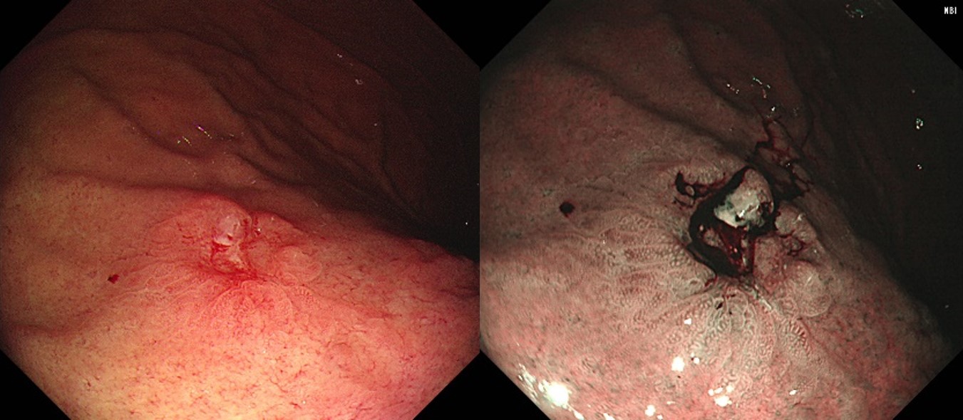
아래는 IIa + IIc와 비슷하기 때문에 조금 깊어보여서 수술과 ESD 사이에서 고민하다가 ESD를 하였던 환자입니다 (위암 292). 침윤깊이는 lamina propria로 생각보다 깊지 않았습니다. 그럼에도 불구하고 약간 융기되어 보였던 것은 GCP 때문인 것으로 추정하였습니다.
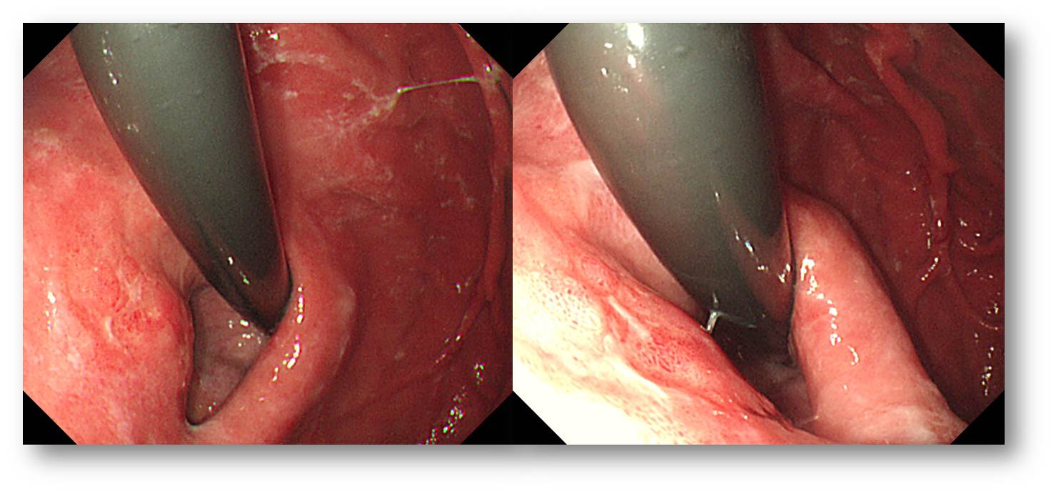
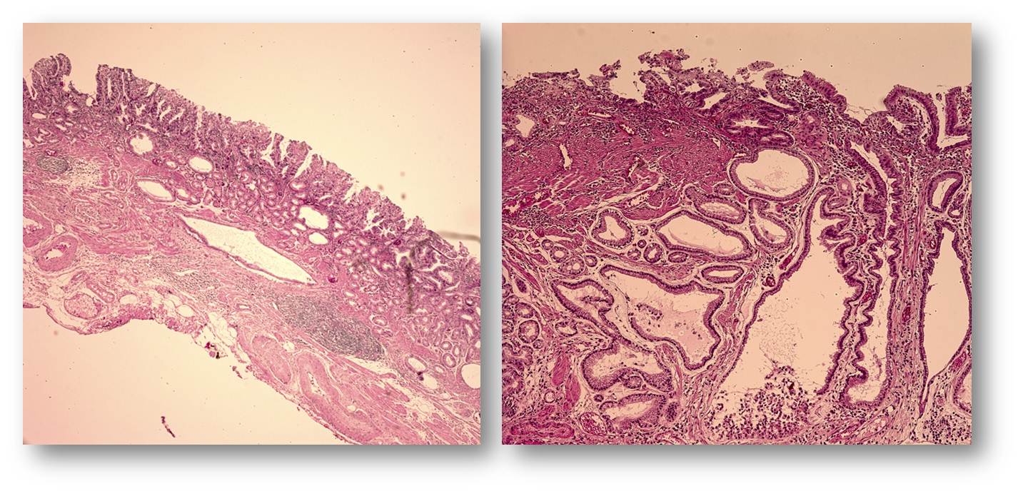
Early gastric carcinoma
1. Location : cardia, lesser curvature
2. Gross type : EGC type IIc
3. Histologic type : tubular adenocarcinoma, well differentiated
4. Histologic type by Lauren : intestinal
5. Size of carcinoma : (1) longest diameter, 20mm (2) vertical diameter, 15mm
6. Depth of invasion : invades mucosa (lamina propria) (pT1a)
7. Resection margin : free from carcinoma(N), safety margin : distal 10 mm, proximal 3 mm, anterior 12 mm, posterior 6 mm
8. Lymphatic invasion : not identified(N)
9. Venous invasion : not identified(N)
10. Perineural invasion : not identified(N)
11. Microscopic ulcer : absent
12. Histologic heterogeneity: absent
13. Associated findings: gastritis cystica profunda
아래도 흥미로운 증례입니다. 2 cm 크기의 위 SMT가 발견되어 시술 전 조직검사에서 암으로 나오지 않은 상태로 의뢰된 환자입니다. Wedge resection을 시행하였고 놀라운 결과가 나왔습니다. 조직검사 위치를 바꾸어 보았다면 수술 전 위암을 진단할 수 있지 않았을까 생각해 보았습니다. 여하튼 herniated gastric mucosa로 인하여 SMT처럼 융기된 부위의 점막 top에서 발생한 위암이라고 결론지었습니다. 혹시 위암이 먼저이고 이로 인하여 gastric mucosa의 herniation이 발생한 것은 아닐까도 생각해 보았습니다.
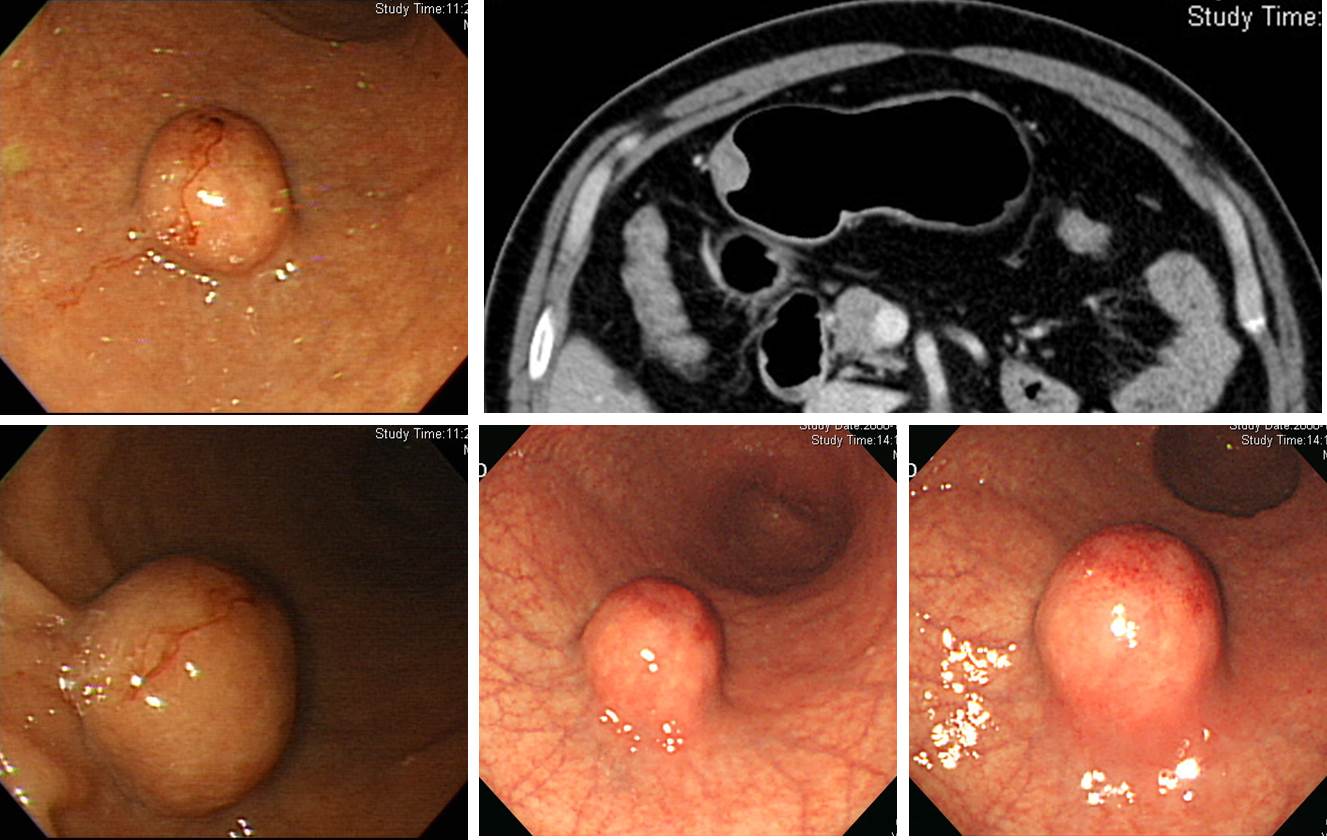
- Histologic type : tubular adenocarcinoma, well differentiated
- Size : 1.5x1.5x1.0 cm
- Depth of invasion : extension to mucosa (muscularis mucosa) (pT1a)
- Resection margin: free from carcinoma
- Associated findings : herniated gastric mucosa in the submucosa
선종과 병발한 경우도 있습니다.
(위질환내시경아틀라스 14호에 실렸던 예수병원 증례)
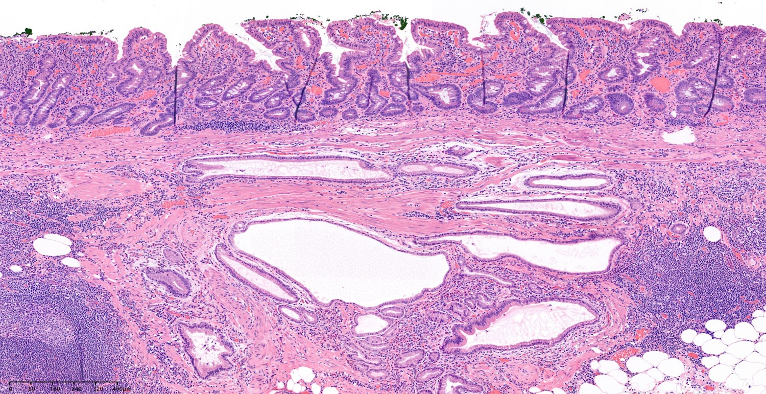
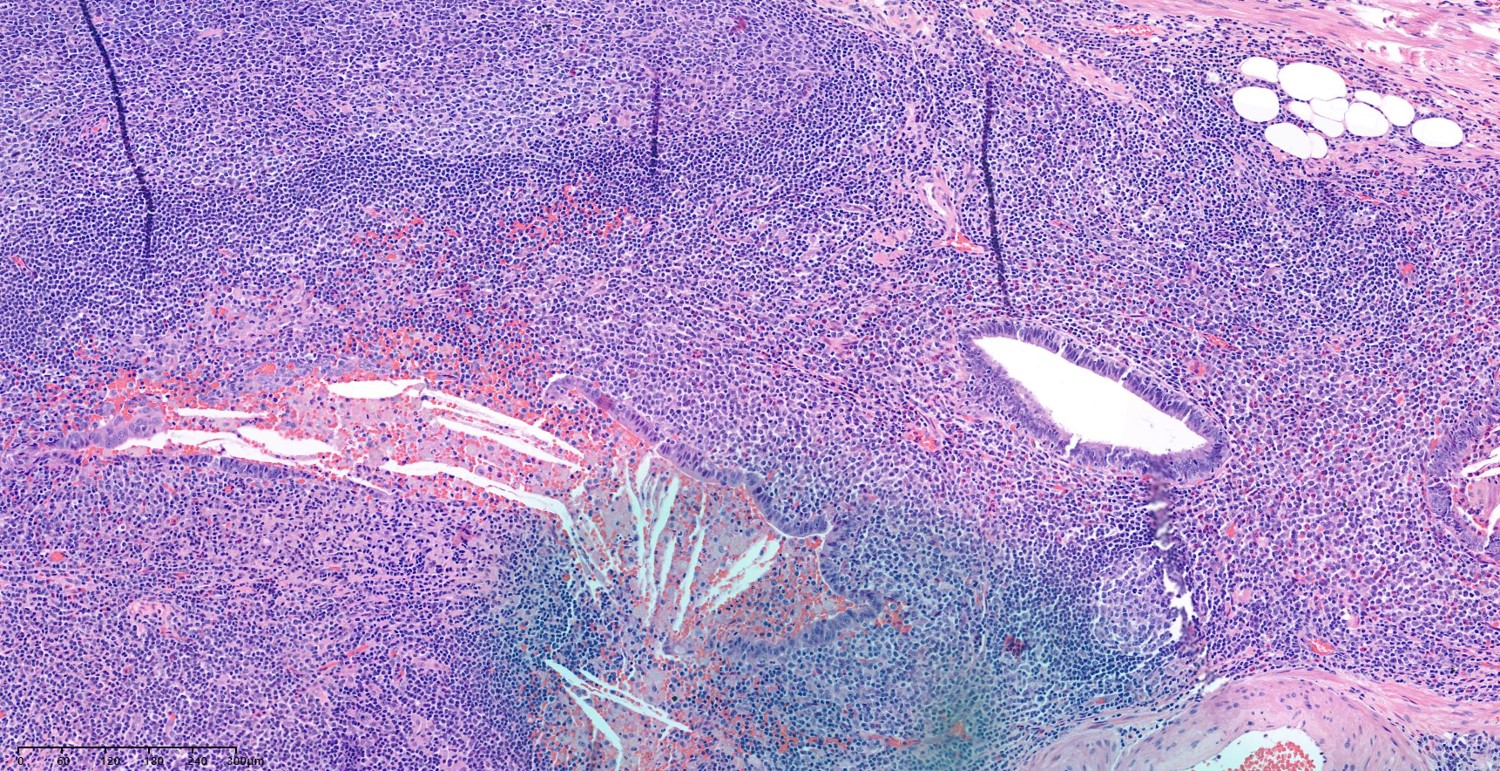
This is a rare mucosal cancer with submucosal gastritis cystica profunda with cancer invasion in the cyst mucosa.
Female 52 (2024)
3. Gastritis cystica superficialis
[References]
1)
© 일원내시경교실 바른내시경연구소 이준행. EndoTODAY Endoscopy Learning Center. Lee Jun Haeng.