EndoTODAY 내시경 교실
Beginner | ESA | Schedule | OPD
Seminars | Atlas | Recent | Links
![]() [외부압박. Stomach extrinsic compression]
[외부압박. Stomach extrinsic compression]
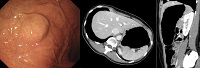
1. 교훈적인 3 증례
2. 비장에 의한 압박 Extrinsic compression by normal spleen or splenic lesions
3. 간에 의한 압박 Extrinsic compression by liver or hepatic lesions
4. 담낭에 의한 압박 Extrinsic compression by gallbladder
5. 다른 장기에 의한 압박 Extrinsic compression by other lesions
6. FAQs
![]() 1. 교훈적인 3 증례
1. 교훈적인 3 증례
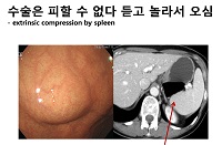
Extrinsic compression은 검진 내시경 후 불필요한 추가 검사 혹은 수술로 연결되기 쉽습니다. 과잉 진단과 과잉 치료가 문제라는 것입니다. 중요한 것은 간혹 진짜 환자가 숨어있다는 것인데요... 그래서 저는 extrinsic compression으로 의뢰되면, 대부분 CT로 확인하고 있습니다. EUS는 워낙 주관적이고 잘못된 판단으로 연결되는 경우가 많아서 좋아하질 않습니다. 교훈적인 증례 세 개를 소개합니다.
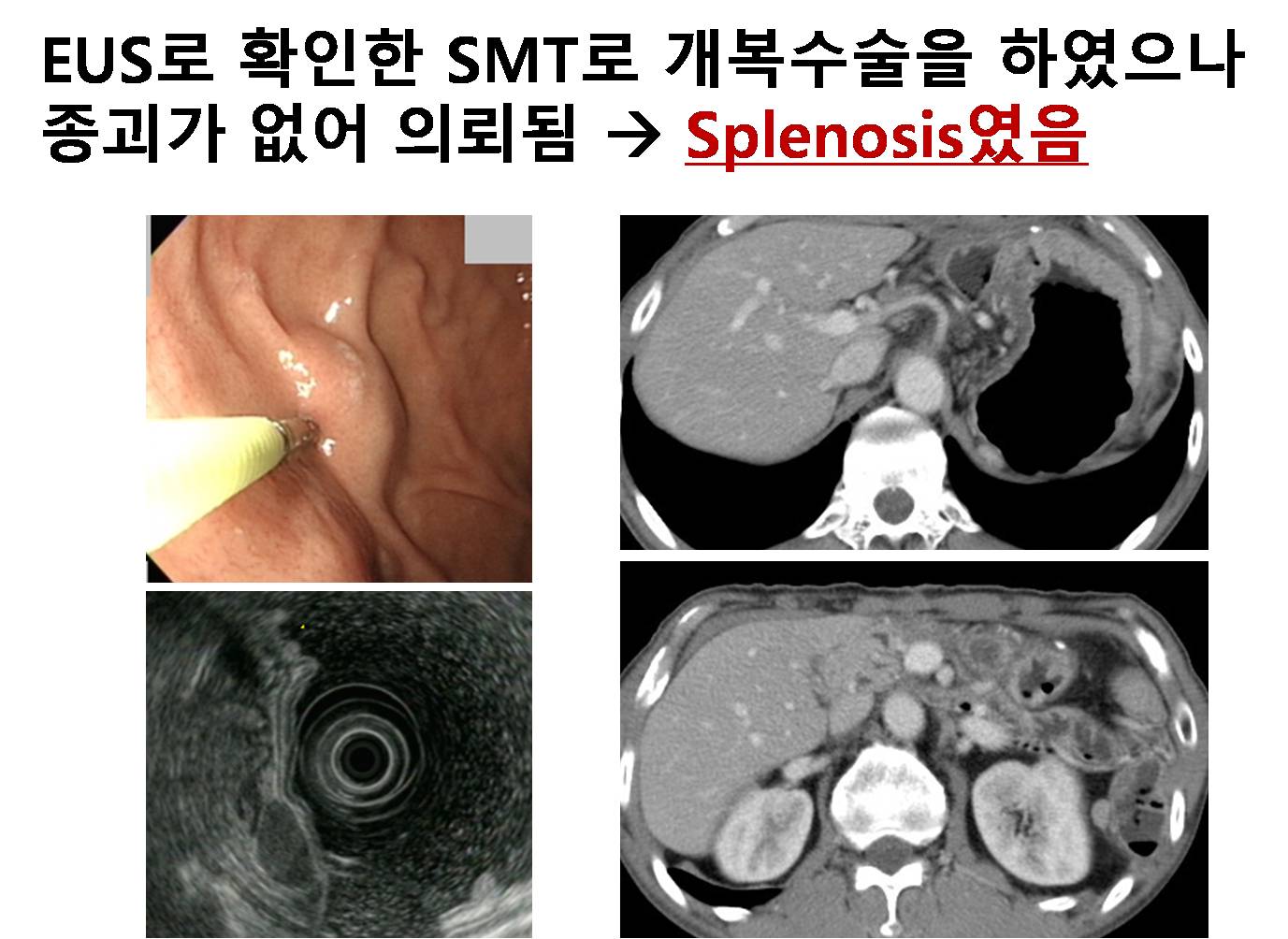
[교훈 증례 1]
SMT라고 EUS를 한 후 수술을 하였는데 막상 SMT가 없어서 의료분쟁으로 연결된 환자입니다. Second opinion을 위하여 저를 찾아오셨습니다. Splenosis였습니다. EUS 소견만 믿고 수술하지 맙시다. 적어도 CT 정도는 확인한 후 수술을 권하기 바랍니다.
이런 증례를 경험하고 나면 EUS에 대한 신뢰감이 확~~~ 떨어집니다.
[교훈 증례 2]
SMT면 자동으로 EUS를 해야 한다고 생각하는 분들이 많습니다. 아닙니다. 위전정부 SMT로 의뢰되었으나 체중감소와 구역감도 있어서 즉시 CT를 시행하여 huge cholangiocarcinoma 진단하였습니다. 무증상 성인의 검진(screening) 내시경인지, 뭔가의 증상이 있어 시행하는 workup 내시경인지 잘 구분합시다. 항상 증상을 물어본 후 검사하는 습관을 가집시다.
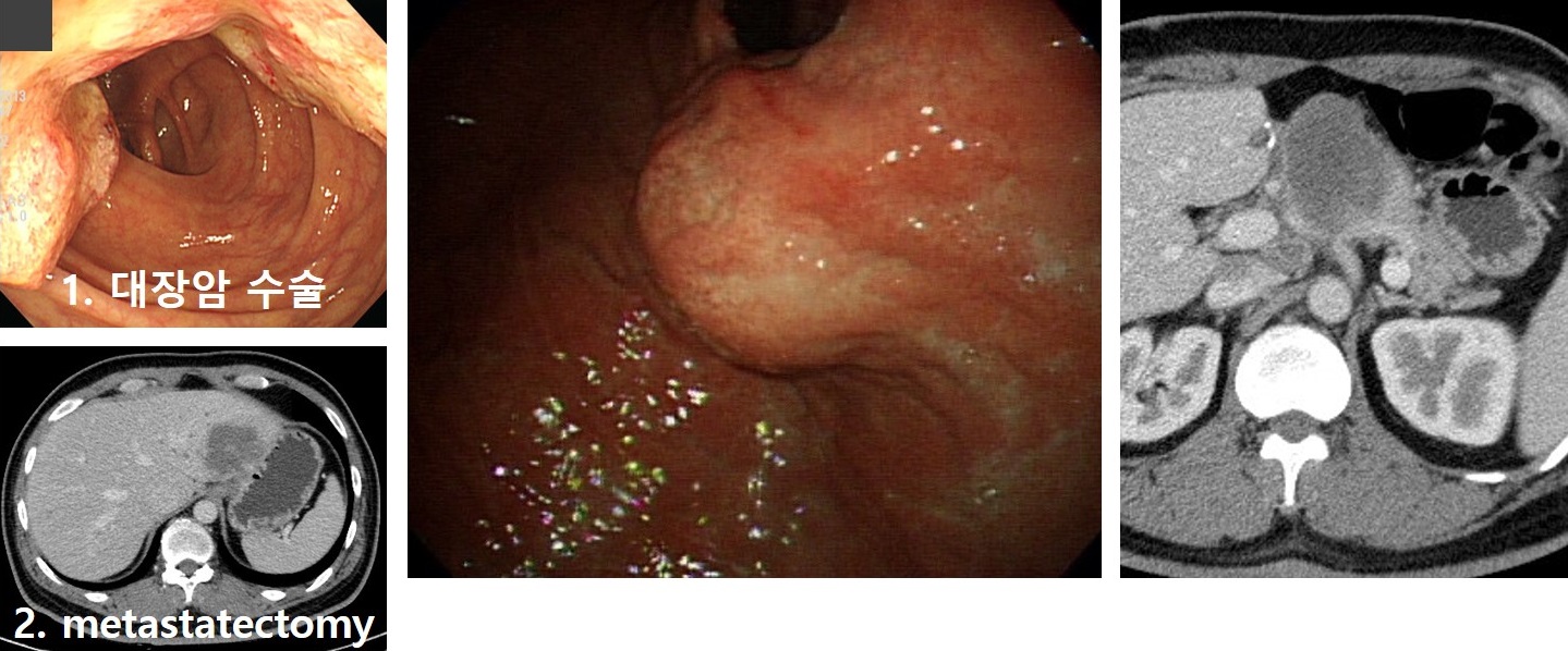
[교훈 증례 3]
Extrinsic compression이 늘 가벼운 병은 아닙니다. 대장암 간전이로 대장수술과 전이수술을 받으신 분이 수년 후 local recur를 보이셨습니다. 내시경 소견은 SMT 혹은 extrinsic compression처럼 보였는데 CT에서는 재발암이 확연하였습니다.
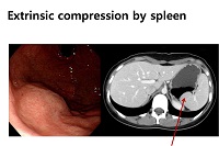
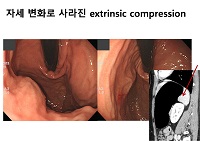
![]() 2. Extrinsic compression by normal spleen or splenic lesions - 비장에 의해서는 fundus나 위체상후 후벽이 눌립니다.
2. Extrinsic compression by normal spleen or splenic lesions - 비장에 의해서는 fundus나 위체상후 후벽이 눌립니다.
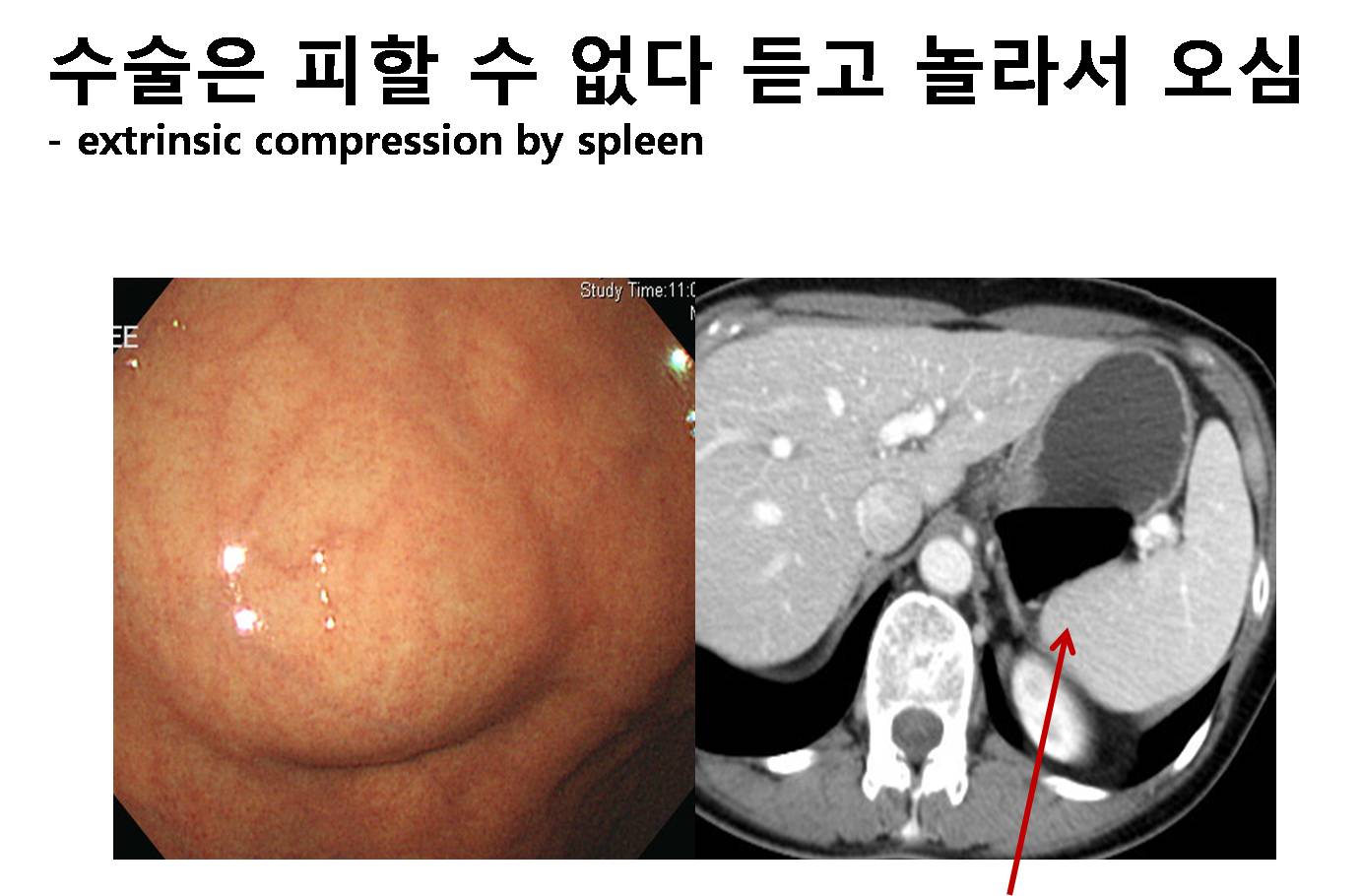
비장의 모양은 참 다양합니다. 위를 누르는 모양도 가지각색입니다.
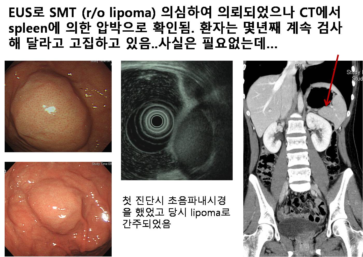
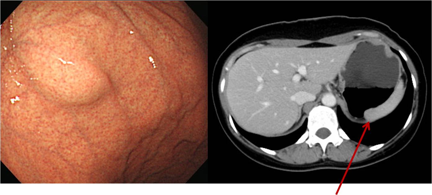
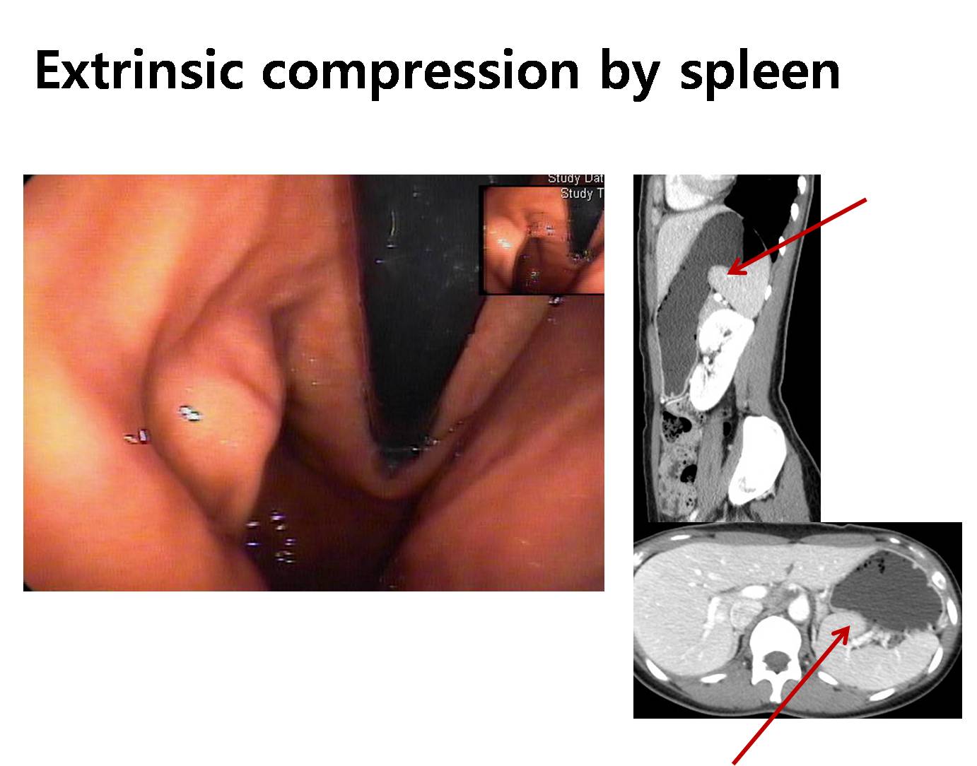
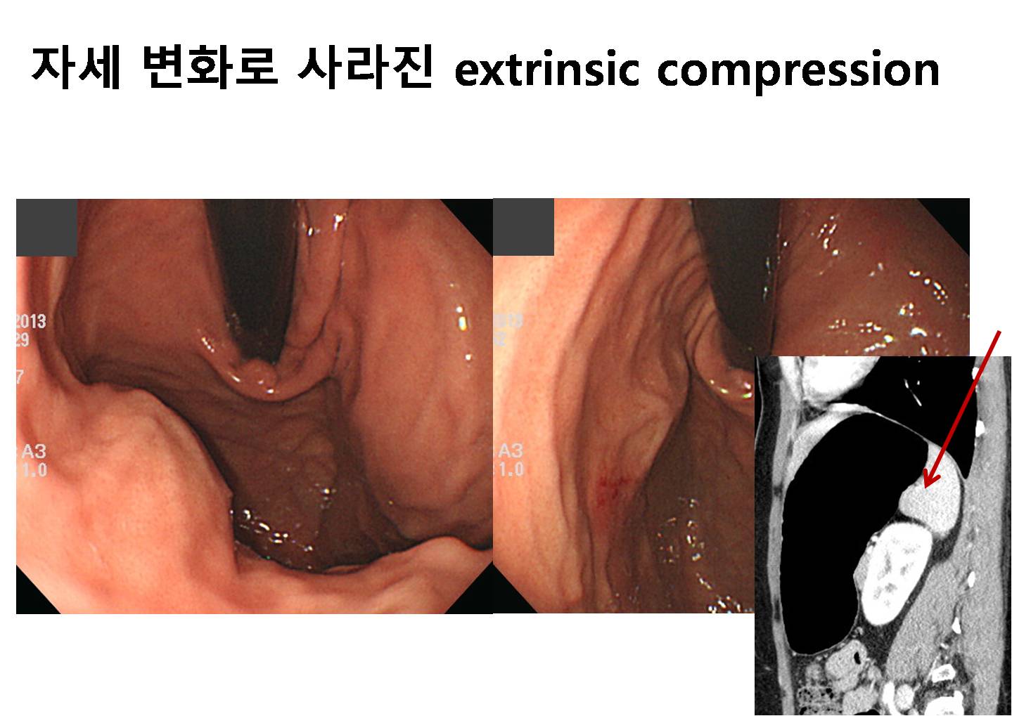
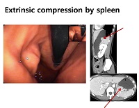
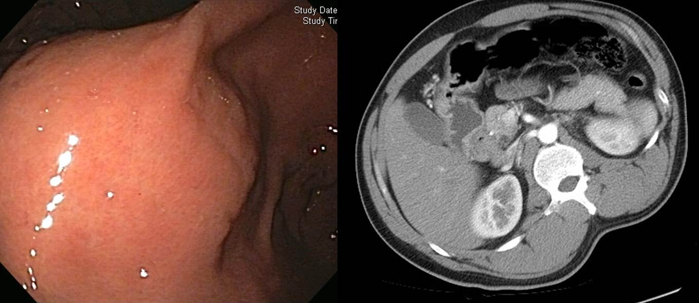
검진 내시경에서 extrinsic compression r/o SMT로 의뢰되었습니다. 사진의 시간을 잘 보면, 처음에는 아무 것도 보이지 않다가 공기를 넣었더니 융기된 부위가 보였고 다시 공기를 빼니 외부 압박처럼 관찰되었던 것이 잘 기록되어 있습니다. 마침 같은 날 찍은 저선량 흉부 CT의 상복부 사진에서 spleen이 위를 누르기 쉬운 모양이었습니다. 이 환자에서 추가 검사가 필요할지 늘 고민입니다. 일단 추가 검사를 위하여 의뢰되면 검사를 하지 않을 수 없는데, stomach CT를 해야할지 EUS를 해야할지 또 다시 고민입니다. 아래 증례들을 참고하여 검사를 한다면 stomach CT를 하는 편이 어떨까 생각합니다.


Screening endoscopy에서 발견된 fundus의 SMT 혹은 extrinsic compression으로 의뢰된 분입니다. 이 경우 follow-up endoscopy, CT 혹은 EUS 중 선택할 수 있습니다. 저는 CT를 선호하는 편입니다.CT에서 위벽의 종괴는 관찰되지 않았고 spleen에 의한 위벽의 indentation이 보였습니다. 내시경 소견을 설명할 수 있다고 판단하고 workup을 중단하고 1년 후 추적내시경만을 권하였습니다. 위 CT는 생각보다 많은 information을 주는 검사입니다. 제가 두번째로 좋아하는 검사입니다. 첫 번째는 당연히 내시경입니다.
Splenic artery aneurysm에 의한 외부 압박
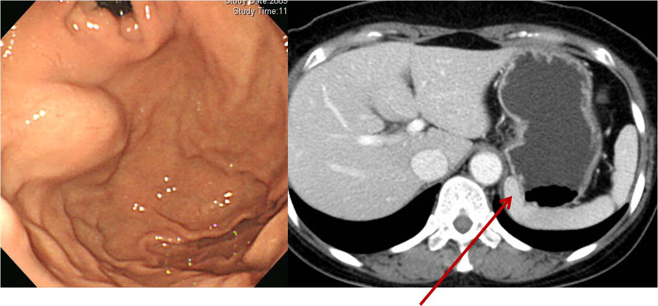
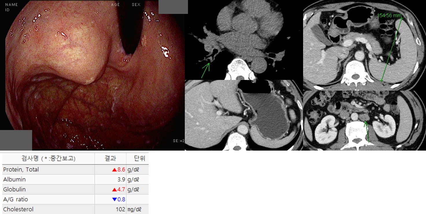
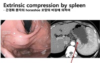
[Gastric extrinsic compression due to splenomegaly by low grade lymphoma]
70 years old female was referred due to gastric SMT-like lesion. On the high body posterior wall, just below the cardia, a 3cm sized protruded lesion was seen. Extrinsic compression due to spleen was suspected. In the CT, splenomegaly, retroperitoneal lymph nodes were found. In the blood chemistry, total protein and globulin were elevated.
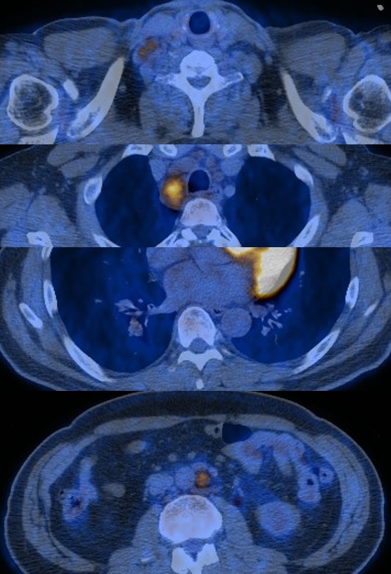
In the PET, hyperemetablic lymph nodes in the right supraclavicular, bilateral mediastinal, both pulmonary hilar, upper abdominal, and retroperitoneal area with splenomegaly were found.
EBUS-transbronchial needle biopsy showed low grade B-cell lymphoma, favoring extranodal marginal zone lymphoma of MALT. Chemotherapy (RCVP) was done and the followup PET and blood chemistry were normalized.
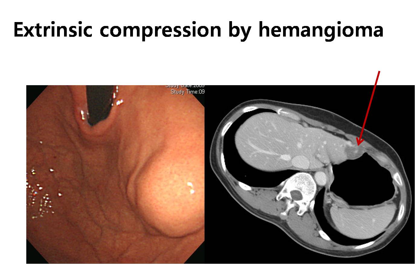
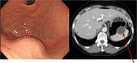
![]() 3. Extrinsic compression by liver or hepatic lesions - 간에 의해서는 주로 전벽쪽이 눌립니다.
3. Extrinsic compression by liver or hepatic lesions - 간에 의해서는 주로 전벽쪽이 눌립니다.
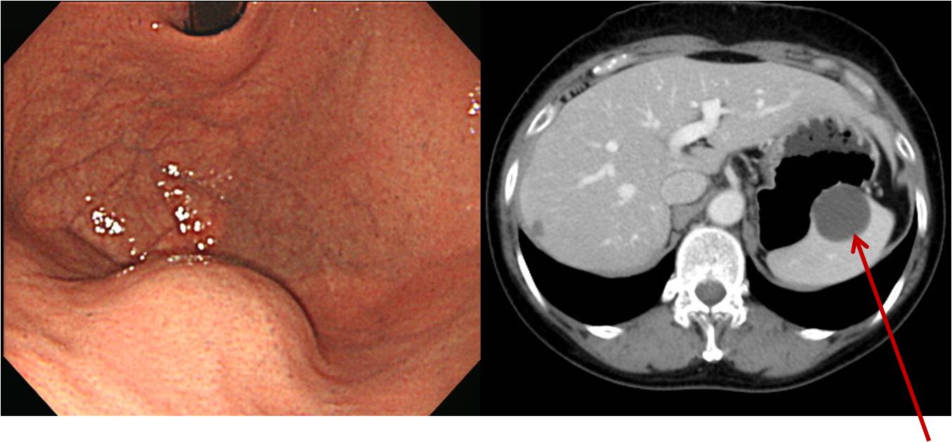
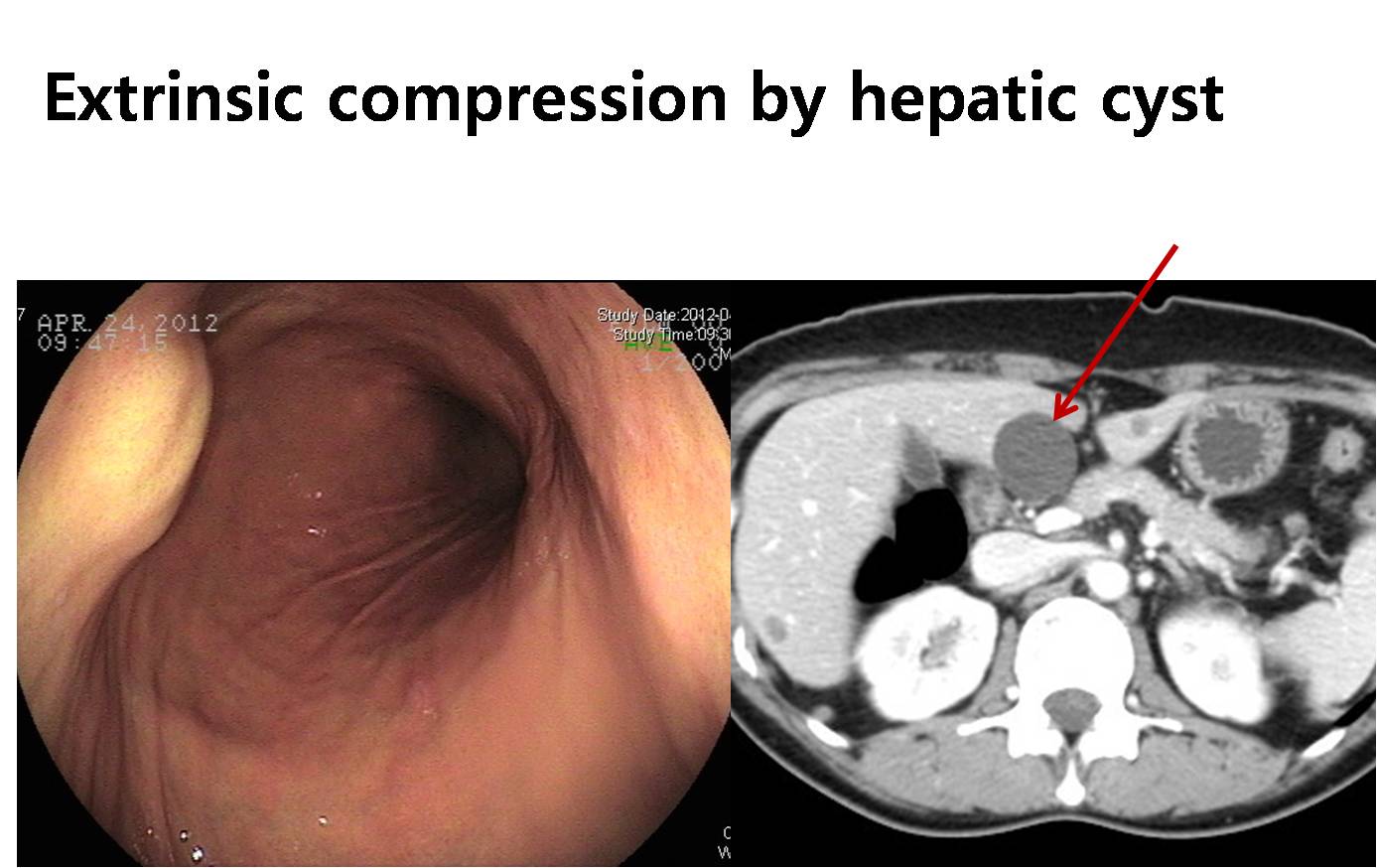
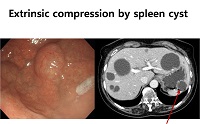
점막하종양이 두 개 있다고 왔는데 하나는 지방종 다른 하나는 간 낭종에 의한 extrinsic compression
간이 푸르스름하게 비쳐보이고 그 중앙에 약간 pale한 small dome-like elevation이 있습니다. 간낭종에 의한 위체상부 전벽 압박의 전형적인 소견입니다.

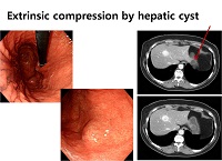
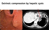

간낭종
간낭종
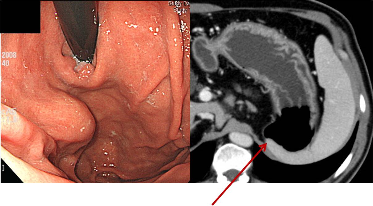
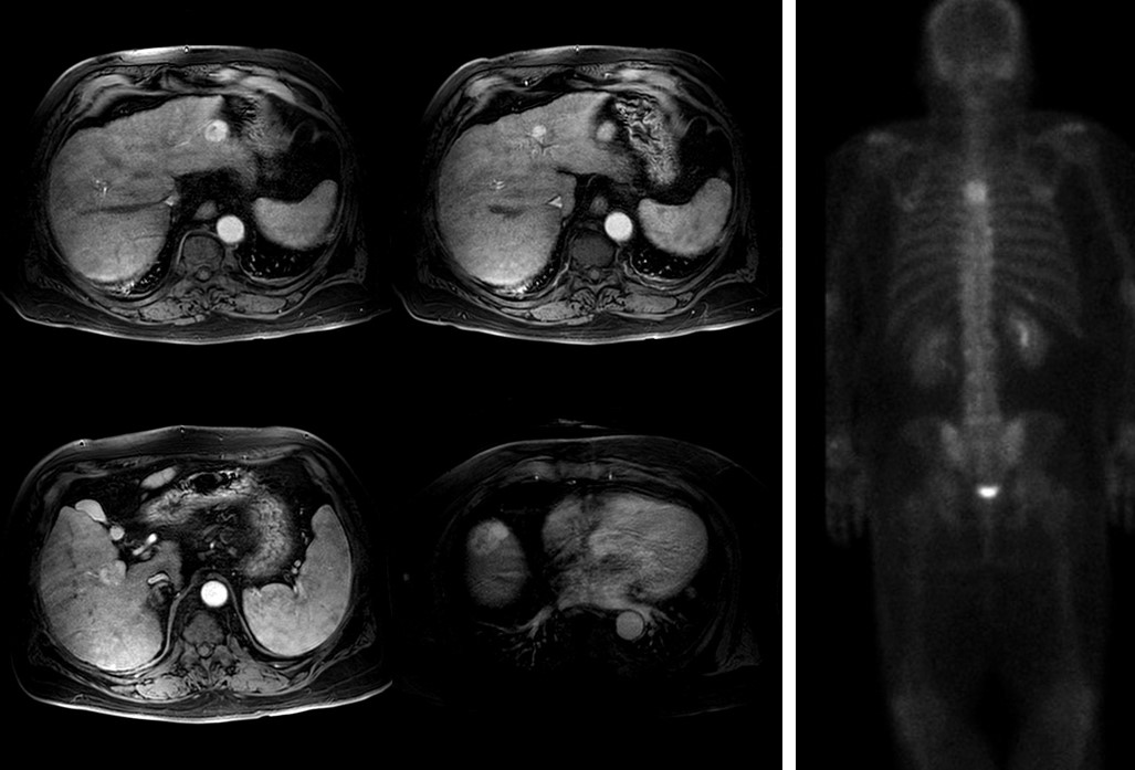
[Extrinsic compression by HCC]
저는 위 SMT에 대한 EUS 검사를 거의 하지 않습니다. 안 한지 몇 년 되었습니다. 과거에는 가끔 하기도 했었는데요... 제가 EUS를 해도 스스로 결과를 믿지 못하는 경우가 많았습니다. 그래서 최근 몇 년 동안은 대부분 stomach CT로 검사하고 있습니다. 아래는 충격적인 증례입니다. 돌이켜 생각해보아도 제가 EUS를 해서 정확히 진단해 낼 수 있었을까 싶습니다. EUS 대신 CT 하기를 잘했다고 생각합니다.
매일 소주를 마셨던 분입니다. 외부 내시경에서 SMT가 발견되어 의뢰되었습니다. Rolling (+), cushion (-)로 씌여 있었습니다. 10일 전부터는 등도 아팠다고 했습니다. 별로 대수롭지 않게 생각하고 CT와 정형외과 의뢰를 했습니다. 그런데, CT 판독이 "(1) Probable HCC in the liver lateral segment. (2) Liver cirrhosis with splenomegaly and prominent splenorenal shunt. (3) No definite evidence of gastric SMT on CT. Probable extrinsic compression due to hepatie lesion. Recommendation: MRI"였습니다.
MRI에서는 multiple HCC가 발견되었고, 혈액검사에서는 HBsAg (-), HCV (-), aFP 145 ng/ml이었습니다. Bone scan에서는 T4 metastasis가 의심되었습니다.
위 SMT에서는 항상 extrinsic compression의 가능성을 고려해야 합니다. 저는 CT를 사랑합니다.
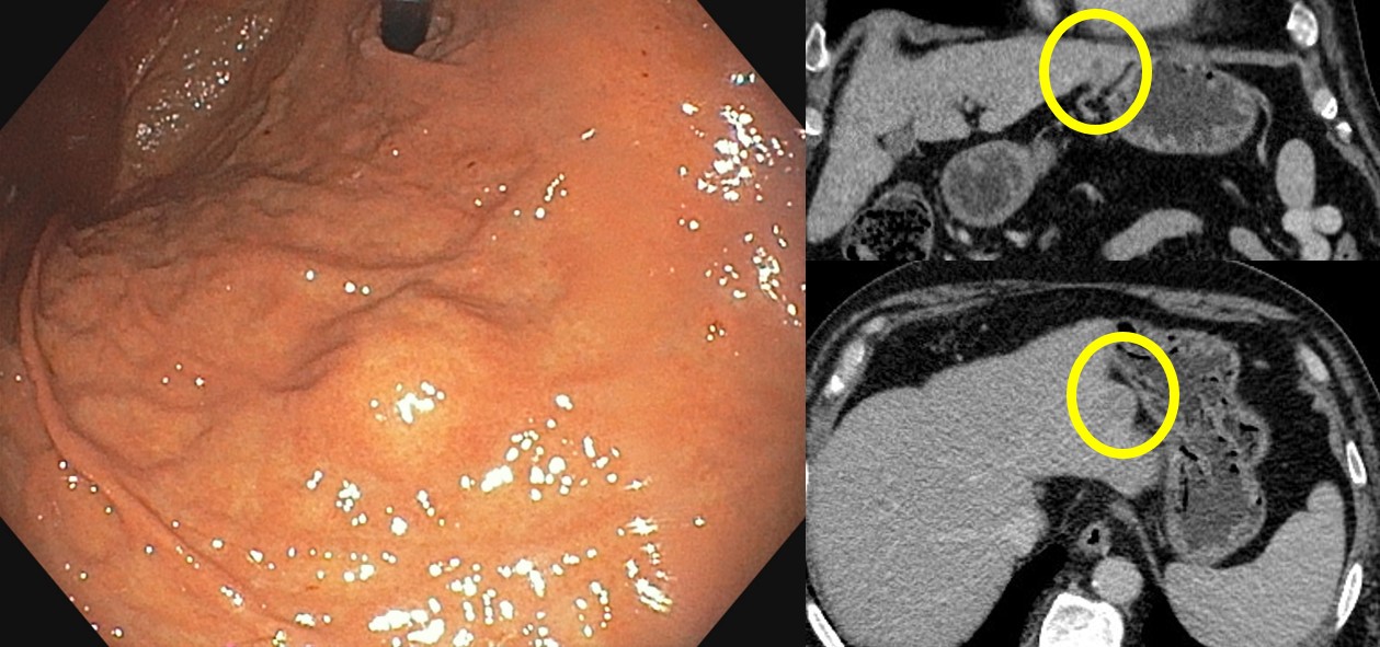
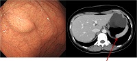
![]() 4. Extrinsic compression by gallbladder
4. Extrinsic compression by gallbladder
2015년. 50대 여성
![]() 5. Extrinsic compression by other lesions
5. Extrinsic compression by other lesions
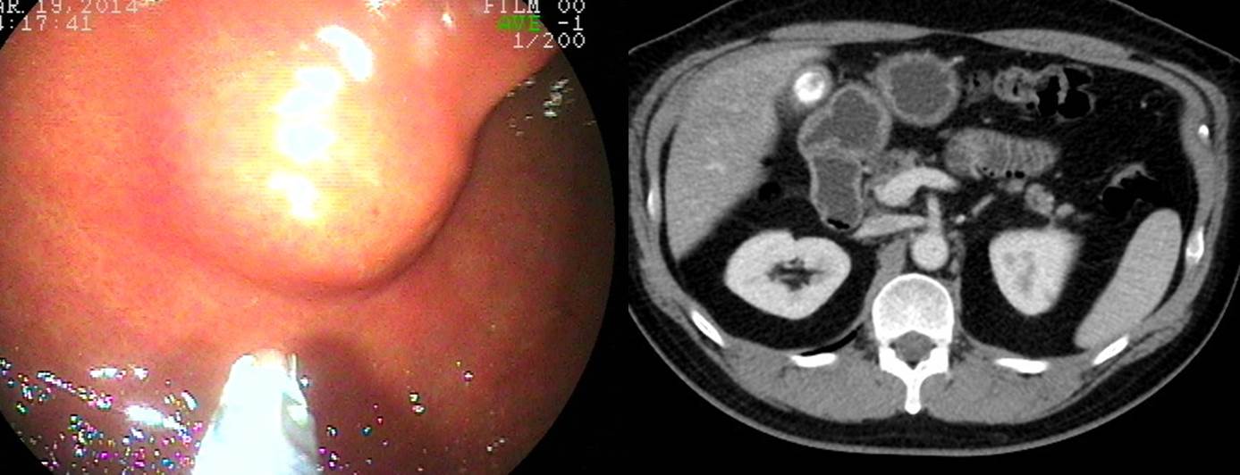
췌장 낭종에 의한 외부 압박
췌장 낭종에 의한 외부 압박
Extrinsic compression due to splenorenal shunt (CT 판독: Gastric body 뒤쪽에 splenorenal shunt를 형성하는 venous structure가 커져 있으며 이것이 위를 누르고 있는 모습)
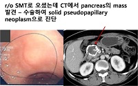
8개월 전 개인 의원에서 위에 혹이 있어 추적관찰을 권유받은 환자입니다. 1개월 전 발생한 복부 불편감으로 검사한 CT에서 pancreas mass with multiple lymph node metastasis and peritoneal seeding 소견으로 의뢰되었습니다. 위 SMT를 가진 환자에서 추적관찰 중 다른 질환이 발생한 예입니다. 위 SMT가 워낙 흔한 것인지라 이러한 환자가 적지 않습니다. 의학적으로 두 질환은 서로 상관이 없지만 환자들은 자꾸 두 질환을 하나로 묶어서 생각하는 경향이 있습니다. 자세한 설명이 필요한 경우라고 하겠습니다.
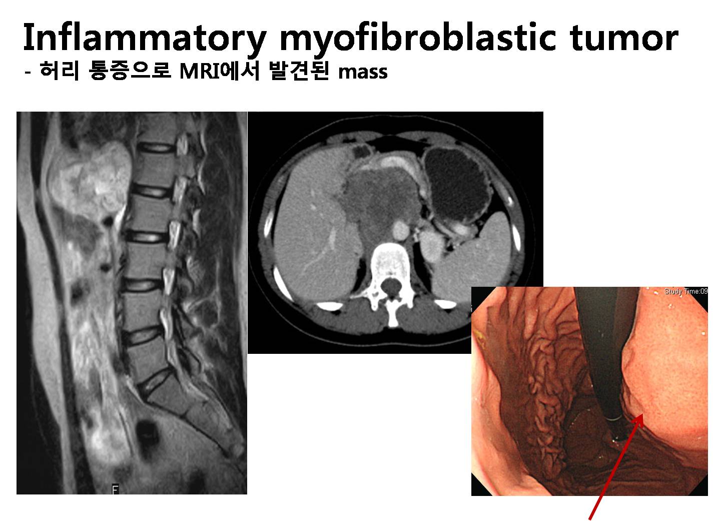
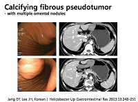
20대 여성이 허리 통증으로 시행한 spine MRI에서 mass가 발견되었습니다 (MRI 판독: L1-2 level의 aortocaval area로 7.7 x 6.3 cm의 solid mass로 생각되는 retroperitoneal mass). 저는 lymphoma의 가능성이 크지 않나 생각하였지만 stomach CT에서는 “Primary retroperitoneal mass such as neurogenic tumor, more likely”라는 impression과 함께 mass의 T2 signal이 높고 불균질하며 내부에 calcification이 있는 점은 전형적인 lymphoma와는 맞지 않는 소견이라는 의견을 얻었습니다. 내시경에서는 위체부의 extrinsic compression으로 관찰되었습니다. 수술을 시행하였고 inflammatory myofibroblastic tumor (IMT)라는 최종 진단을 받았습니다. IMT는 intermediate biologic potential을 가지는 neoplasm으로서 저는 처음 경험하기 때문에 참고 문헌의 일부를 소개하는 것으로 설명을 대신하겠습니다.
Coffin CM, et al. Semin Diagn Pathol 1998;15:85-101. IMT or inflammatory pseudotumor was initially recognized in the lung, and somewhat later, a similar-appearing pathological process was reported in the liver. Presently, this tumor has been described in virtually all major organs and extrapulmonary sites with a few exceptions. It was thought initially that the IMT was nonneoplastic and represented an aberrant inflammatory response despite its gross and microscopic features of a spindle cell neoplasm. The inflammatory hypothesis about the pathogenesis has been more readily accommodated in the lung than in the extrapulmonary sites of involvement. Some cases, however, were accompanied by the constitutional symptoms and signs of an inflammatory process, which resolved in most cases after surgical resection. There were some pathological aspects of the IMT that seemingly contradicted its purely inflammatory nature, including its potential for local recurrence; development of multifocal, noncontiguous tumors; infiltrative local growth; vascular invasion; and malignant transformation. These pathological features seemed to support the hypothesis that the IMT is a neoplastic process, which has been augmented by reports that these tumors have clonal characteristics. Because these tumors have a predilection for children, embryonal rhabdomyosarcoma is another diagnostic temptation when an IMT presents in the bladder or other hollow viscus.
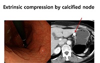
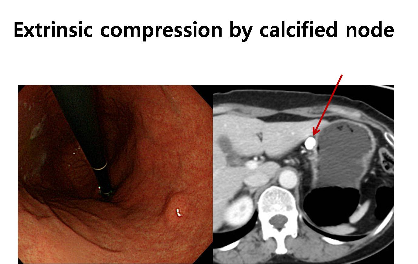
Extrinsic compression by calcified splenic aneurysm. 내과의사의 눈으로는 calcified lymph node와 구분이 어려웠습니다.
위전정부 SMT로 의뢰되었으나 체중감소와 구역감도 있어서 즉시 CT를 시행하여 huge cholangiocarcinoma 진단
![]() [FAQ]
[FAQ]
[2014-7-26. 애독자 질문]
저는 검진센터에서 일하고 있습니다. 평소 교수님이 보내주시는 자료로 열심히 공부하고 있습니다. 내시경을 하면 할수록 모르는게 생기는것 같습니다. 위내시경상 정상적으로 외부장기에 의해 압박이 있을수 있는데 정상과 비정상의 구분이 어려운 경우가 있습니다. 20세 여자환자가 검진 내시경을 했는데 위저부에 아래 그림과 같은 병변을 보였습니다. 정상적인 외부장기에 의한 압박인지 아니면 상피하종양이 있는건지 여쭤보고 싶고 이런경우 꼭 복부CT를 찍어야 하는지 여쭤보고 싶습니다.
[2014-7-31. 이준행 답변]
SMT는 과잉진단이 난무하는 영역입니다. 특히 extrinsic compression으로 불필요한 검사를 하거나 경우에 따라서는 수술까지 하고 난 후 아무 것도 없다고 듣는 환자까지 있습니다. 증례 사진을 많이 보는 수 밖에 없을 것 같습니다. 제가 경험하였던 증례 일부를 소개합니다.
그런데 왜 20세 여자가 건진 내시경을 받지요? 이것부터가 과잉이라고 생각합니다.
© 일원내시경교실 바른내시경연구소 이준행. EndoTODAY Endoscopy Learning Center. Lee Jun Haeng.