 EndoTODAY ���ð� ����
Beginner | ESA | Schedule | OPD
Seminars | Atlas | Recent | Links
![]() [ColonTODAY 041 - Colon amyloidosis]
[ColonTODAY 041 - Colon amyloidosis]
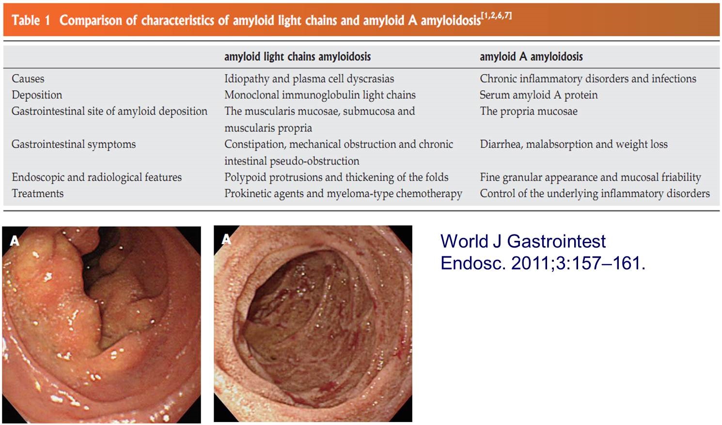
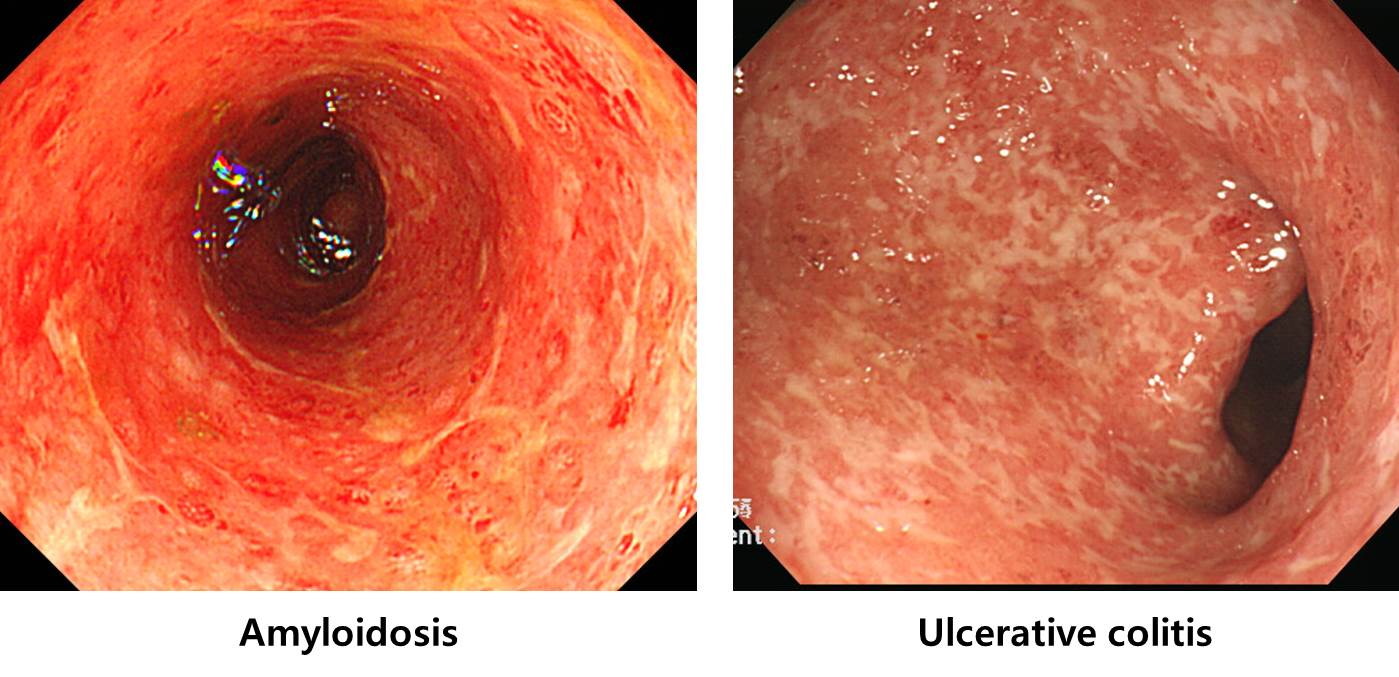
���� amyloidosis�� ulcerative colitis�� ��Ȥ ����� ���� �� �ֽ��ϴ�. ���� ���� ������ �ٸ���... �������� �ӻ���� ���� �ٸ��ϴ�.
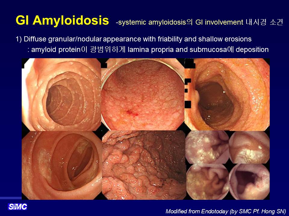
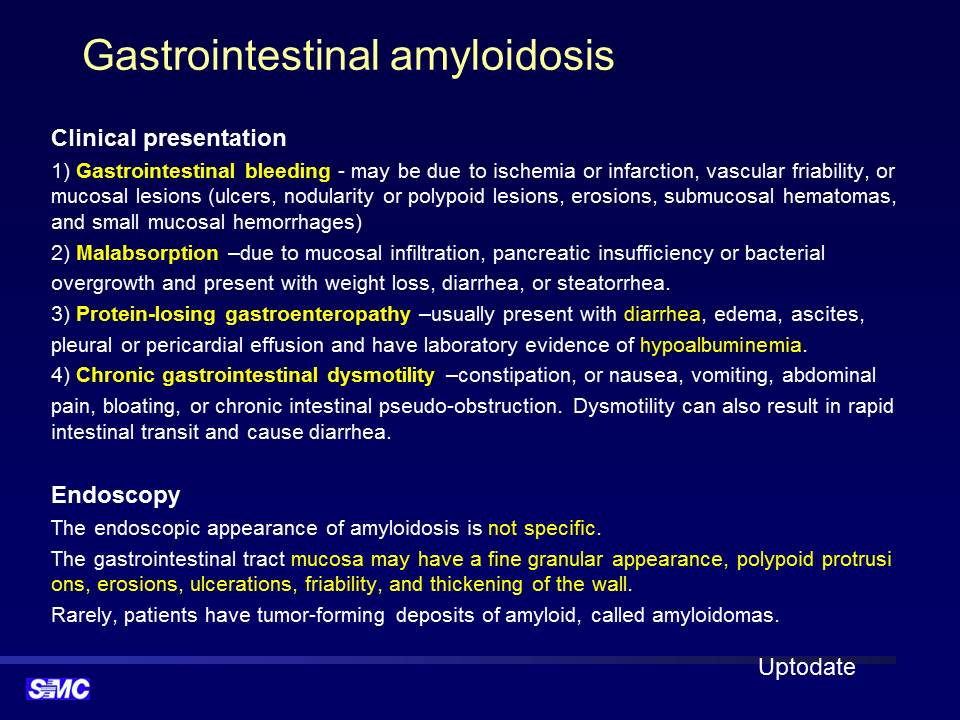
![]() 1. Introduction - �ƹз��̵���
1. Introduction - �ƹз��̵���
�ƹз��̵����� �����ϱ� ����� ���Դϴ�. Primay amyloidosis (multiple myeloma ���� ���ϰ� ���ݵ� ���, AL amyloidosis)�� secondary amyloidosis (����Ƽ�� ������ �� ���� ������ ��ȯ�� ���ݵ� ���, AA amyloidosis) �� ��Ÿ �� ���� ������ �ֽ��ϴ�.
Amyloidosis is a group of infiltrative disorders that result from the extracellular deposit of amyloid fibrils composed of a variety of serum protein precursors, along with the nonfibrillar glycoprotein serum amyloid P (SAP) and glycosaminoglycans. Over 20 different proteins have been identified as causative agents. The letter A is used to designate amyloid fibril protein and is modified by a second letter or letters to indicate the specific fibrillar protein. Thus, with primary amyloidosis, the most common form is called AL, the L representing the fragment of immunoglobulin light chains found in the majority of patients, whether ��primary�� or associated with multiple myeloma. (Sleisenger ������)
Amyloidosis is characterized by extracellular deposition of abnormal protein. There are six types: primary, secondary, hemodialysis-related, hereditary, senile, and localized. Primary (AL) amyloidosis is associated with monoclonal light chains in serum and/or urine with 15% of patients having multiple myeloma. Secondary (AA) amyloidosis is associated with inflammatory, infectious, and neoplastic diseases. The presentation is protean, including macroglossia, a dilated and atonic esophagus, gastric polyps or enlarged folds, and luminal narrowing or ulceration of the colon. Amyloid deposition in the gastrointestinal (GI) tract is greatest in the small intestine. The symptoms include diarrhea, steatorrhea, or constipation. Pseudo-obstruction carries a particularly grave prognosis, often not responding to pro-motility agents. Hepatic involvement is common, but the clinical manifestations are usually mild with hepatomegaly and an elevated alkaline phosphatase level. Biopsies to diagnose amyloidosis can be taken from the fat, kidney, intestine, or bone marrow. The safety of liver biopsies is controversial. With Congo Red stain, amyloid appears red in normal light and apple-green in polarized light. Treatment for AL amyloidosis is chemotherapy and stem cell transplantation; treatment for AA amyloidosis is control of the underlying disease. Amyloidosis should be considered in patients with proteinuria, cardiomyopathy, hepatomegaly (with mildly abnormal liver tests), peripheral and autonomic neuropathy, weight loss, and GI symptoms. (Ebert EC. Am J Gastroenterol 2008)
Systemic amyloidosis�� ������ ���� ��⸦ ħ���� �� �ֽ��ϴ�. �� �� �� amyloidosis�� ó�� ���ܵ� ȯ�ڰ� workup�� ���Ͽ� �Կ� ù �� ����� ��찡 �־����ϴ�. ������ cardiac amyloidosis. ������ ���Դϴ�.
2025�� ����� amyloidosis�� ���ð� �з��� ���ȵǾ����ϴ�. (Gut and Liver 2025)
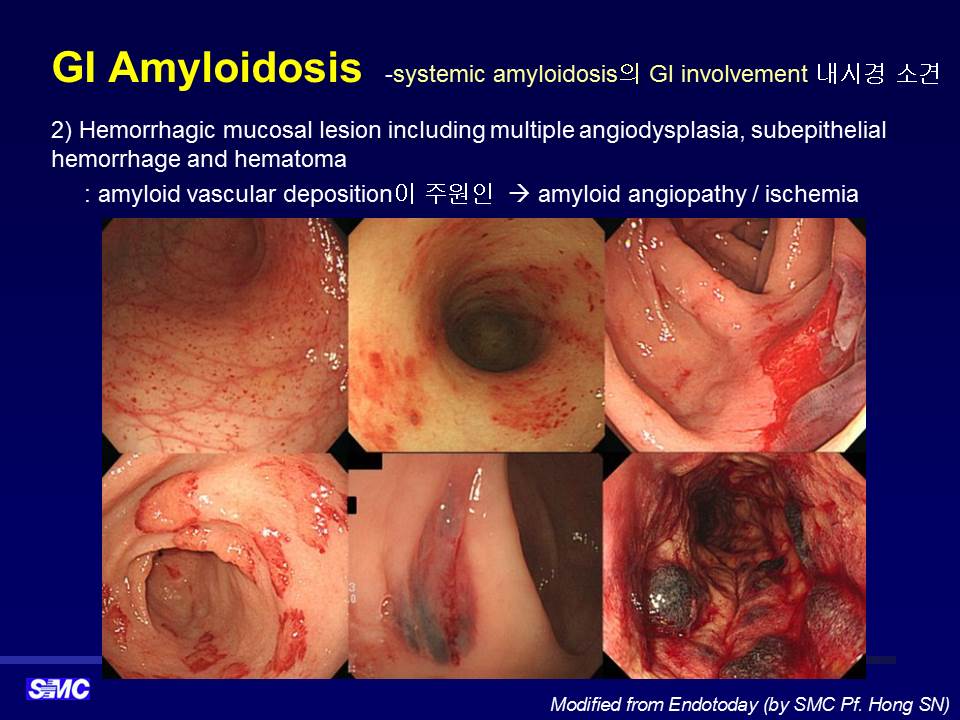
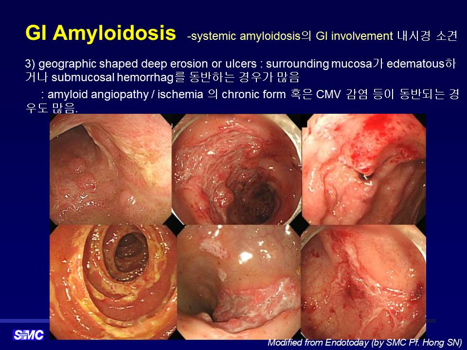
![]() 2. Colonic involvement of the systemic amyloidosis ���� �ƹз��̵����� ���� ħ��
2. Colonic involvement of the systemic amyloidosis ���� �ƹз��̵����� ���� ħ��
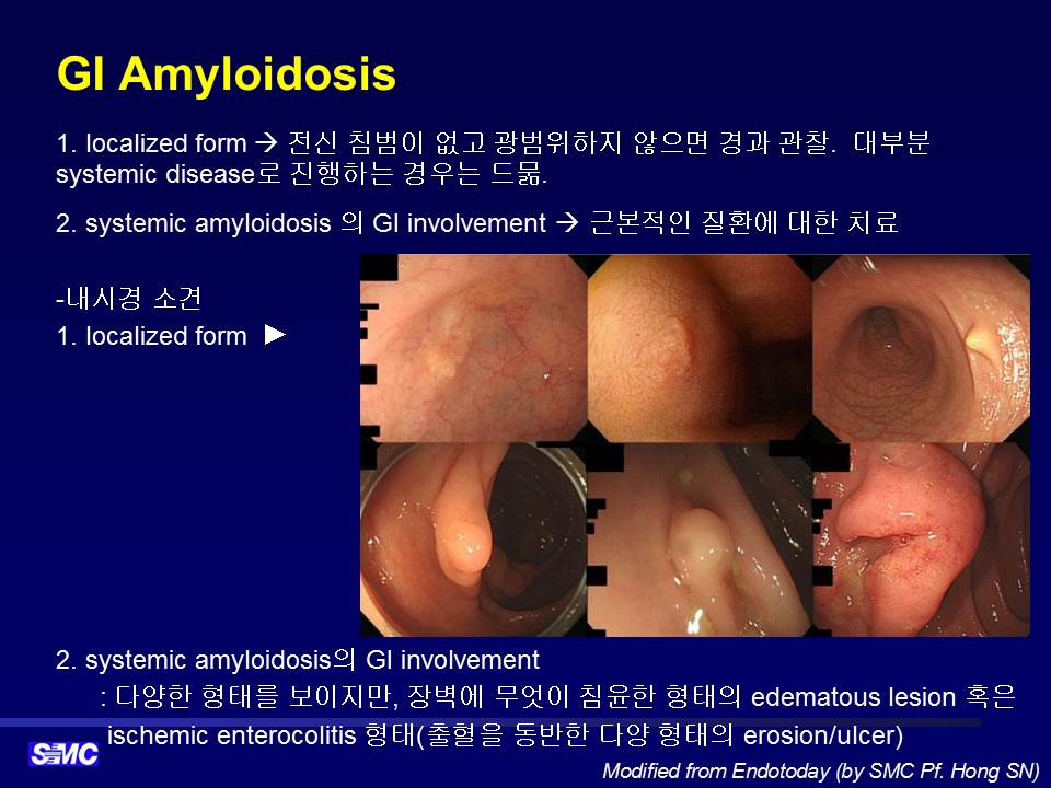
���忡�� �߰ߵ� �ƹз��̵����� ���� �˻縦 �� ���� primay amyloidosis (multiple myeloma ���� ���ϰ� ���ݵ� ���, AL amyloidosis)�� secondary amyloidosis (����Ƽ�� ������ �� ���� ������ ��ȯ�� ���ݵ� ���, AA amyloidosis)�� ���� ���մϴ�. �幰�� localized form�� �����մϴ�.
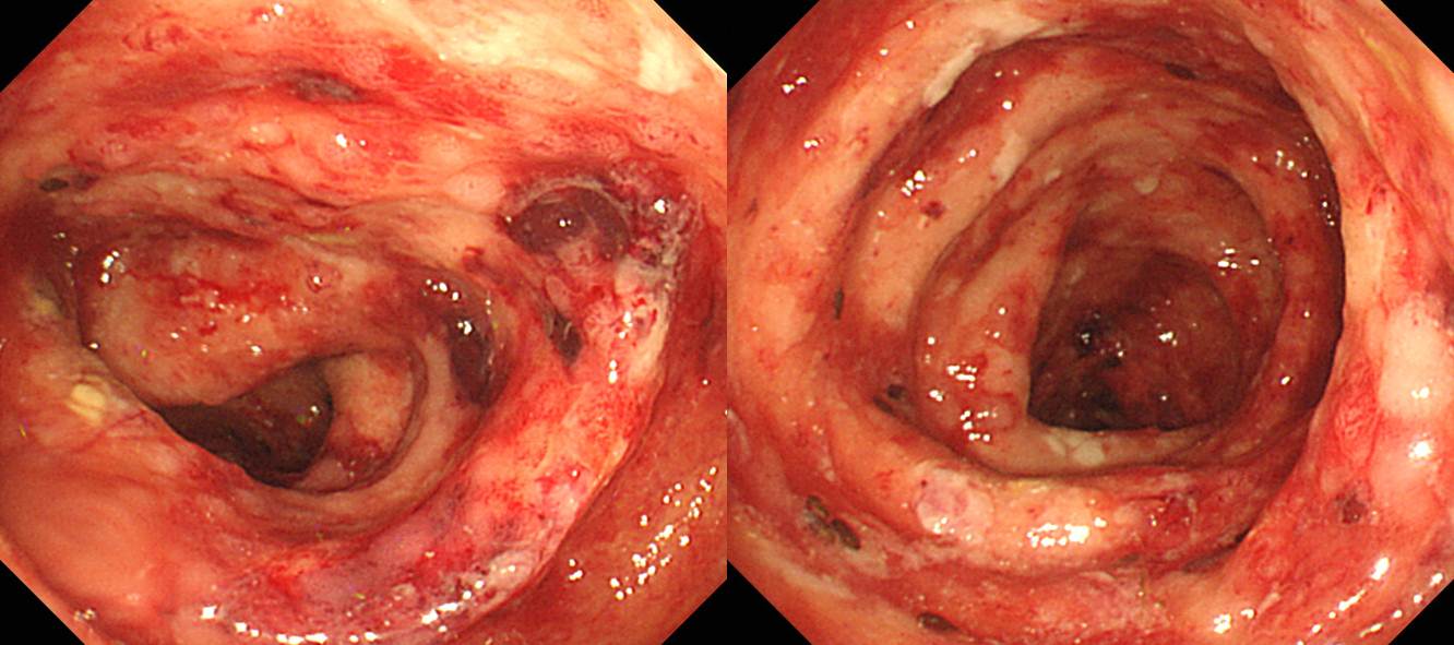
Multiple myeloma ȯ���� ���� ���� amyloidosis
���� lymphoplastmocytic lymphoma (���� 1, ���� 2)�� ġ�� ���� 70�� �������� �������縦 ������ ���� �����˻�� ���� amyloidosis����.
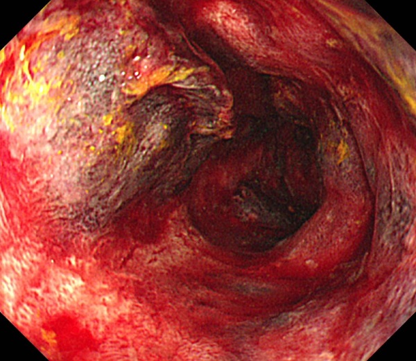
TB destrolyed lung, CNPA (chronic necrotizing pulmonary aspergillosis) ȯ�ڰ� ������ ��ȭ�ҷ�, �����ظ�, poor oral intake�� ���峻�ð� �˻縦 �����Ͽ� �����˻翡�� amyloidosis�� ���ܵǾ��� ��� �˻翡�� plasma cell�� �����Ǿ� �־���, ���װ˻翡�� polyclonal gammopathy�� ����. ���� ������ ���� secondary amyloiodosis (AA type)���� �Ǵ��Ͽ� CNPA�� ���� �������� ���� �� ���� ���� ���� ġ�� ����.
��ü�� ȯ�ڿ��� ���� systemic amyloidosis
![]() 3. Localized amyloidosis
3. Localized amyloidosis
���� ħ���� ���� �ѵ� �� ���ѵ� localizad form�� �ֽ��ϴ�. ���Ĵ� �����ϴ�.
40�� ������ ���� ���峻�ð濡�� �߰ߵ� localized form�� colon amyloidosis�Դϴ�. Ÿ ��� ��ȯ�� �������ϴ�. ��������� �ϴ� �� �ۿ� �����ϴ�. ��� ���Ĵ� ���� �� �����ϴ�.
40�� ����. Hematochezia
Localized amyloidosis ���� �ڷḦ �Ұ��մϴ�.
Localized deposition of amyloid may occur in individual organs, in the absence of systemic involvement. The reason for localized deposition is unknown, but it is hypothesized that deposits result from local synthesis of amyloid protein, rather than the deposition of light chains produced elsewhere. We identified 20 cases of localized amyloidosis at our institution between 1993 and 2003. There were 11 males and nine females in the group. The mean age at the time of diagnosis was 65.5 years. Organs involved included skin, soft tissues, oropharynx, larynx, lung, bladder, colon, conjunctiva, and lymph node. In six of nine patients typed, the amyloid light chain was lambda. In those patients where follow-up was available (mean 7.6 years), none developed systemic disease. Localized amyloidosis occurs in a variety of organ systems. Evolution into systemic amyloidosis was not seen in our series of patients, supporting the hypothesis of local production of amyloid protein in these cases. (Biewend ML. Amyloid 2006)
![]() 4. Secondary amyloidosis ���� - RA ȯ���� chronic diarrhea�� ������ secondary amyloidosis (EndoTODAY ������ɳ��ð�����ȸ 2018-2-22)
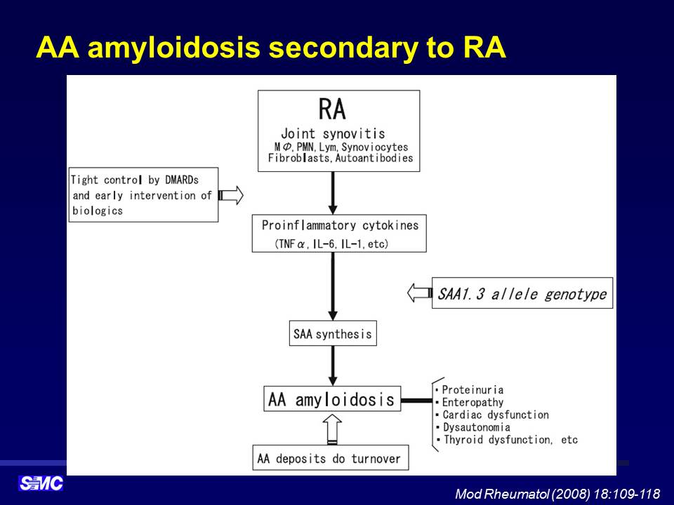
4. Secondary amyloidosis ���� - RA ȯ���� chronic diarrhea�� ������ secondary amyloidosis (EndoTODAY ������ɳ��ð�����ȸ 2018-2-22)
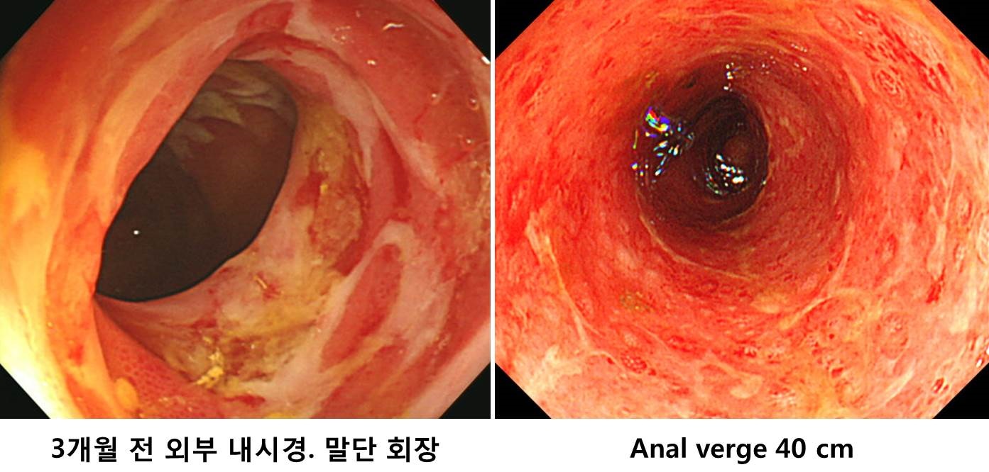
�� ���� �� �ܺ� ���峻�ð濡�� terminal ileum�� diffuse shallow ulcerative lesion�� scattered �Ǿ� �־��� ������ ��� ��Ȯ�� ���������� ������ �ʾҰ�, RA ���� �忰 �ǽ��Ͽ� ���� ġ�� �� ȣ���� ������ �ֽ��ϴ�.
������, �߿��� ����� �Ƿڵ� sigmoidoscopy���� anal verge 30cm ���� 60cm ���� ������ vascular pattern�� �ҽǵǰ� superfical ulcerative lesion�� scattered �Ǿ� �����˻縦 �Ͽ��� amyloidosis�� Ȯ�εǾ����ϴ� (Congo red: positive; amyloid P: positive; TTR: negative; lambda light chain: negative; kappa light chain: negative, amyloid A: positie).
������ ���ؼ��� r/o septic knee�� ������ �����ϸ鼭 ���� RA�� ���� ���뷮 steroid�� �����Ͽ��� ���� ȣ���Ǿ����ϴ�. ���絵 ���������ϴ�. �������系�� �������� "RA ���� AA amyloidosis�̸� GI involvement only ��Ȳ�̹Ƿ� ġ�� ���� 1�� �� follow-up"�� ���ڴٴ� �ǰ��̾����ϴ�.
������ �����Բ��� ª�� ���並 �� �ּ̽��ϴ�.
![]() 5. ����� �ƹз��̵����� ġ��
5. ����� �ƹз��̵����� ġ��
1) Systemic GI amyloidosis�� ����� ħ��: �ٺ����� ��ȯ�� ���� ġ��
2) Localized form: ���� ħ���� ���� ���������� ������ ��� ����. ������ȯ���� �����ϴ� ���� �年�ϴ�.
�Z���ﺴ�� ȫ���� �������� ������ �����Ͻñ� �ٶ��ϴ�.
PDF 0.6 M
![]() 6. ���� �ƹз��̵����� ���� Quiz (EndoTODAY 20150624)
6. ���� �ƹз��̵����� ���� Quiz (EndoTODAY 20150624)
[Quiz]
50�� ���ڷ� �ǰ��������� �ܹ鴢�� �߰ߵǾ����ϴ�. �� ���� �� ����(������Ƹ� ����)�� �Ͽ��� �㸮�� ���� ������ �湮�Ͽ� ô�� �йڰ���, ����, creatinine ����� �߰ߵǾ����ϴ�. �˻縦 �Ͽ��� � �������� ���ܵǾ����ϴ�. ġ�� �� �������� ���峻�ð��� �Ͽ����ϴ�. (1) � ������ �ǽɵǸ�, (2) ���峻�ð��� � �Ұ��̰� ������ �����̰ڽ��ϱ�?
[�亯]
������ �ʹ� ��������? ��Ʈ�� �ʹ� ���ҳ���? ��� �� �¾ҽ��ϴ�. Multiple myeloma with colon amyloidosis�����ϴ�. �� �����о߰� �ƴ����� �Z���ﺴ�� ȫ���� �����Բ� �ؼ��� ��Ź��Ƚ��ϴ�. �ڼ��� ������ �����ּ̽��ϴ�. �����մϴ�. �Ϻθ� �ű�ϴ�. �� PDF file�� ��� ���� �о�ñ� �ٶ��ϴ�.
ȯ���� ���µ��� �����ϸ� amyloidosis �����ϸ�, ������ �ٸ� ������ ������ ������ �پ��� ������ �̶�/�˾��� ȥ���ؼ� infectious/ ischmic colitis�� ���ܵ� �� �ֽ��ϴ�. ���� ȯ���� sigmidoscopy ���� �� �Ʒ��� ���� submucoal hematoma�� �����˴ϴ�.
�� ���������� GI bleeding�� ������ AL amylodosis�� ������ ���ð� �Ұ����� ��ǥ�� �� �ֽ��ϴ�. James DG. Clinical recognition of Al type amyloidosis of the luminal gastrointestinal tract. Clin Gastroenterol Hepatol 2007
RESULTS: Nineteen patients with systemic AL amyloidosis of the luminal gastrointestinal tract were identified. Gastrointestinal symptoms or signs related to amyloid involvement were noted in 95% of patients; abdominal pain, change in bowel habits, overt gastrointestinal bleeding, and complaints related to altered motility were the predominant presentations. Endoscopic abnormalities were found in nearly three fourths of patients, including ulcerations and submucosal hematomas. When gastrointestinal bleeding was the presenting symptom, submucosal hematomas were a common finding during endoscopic evaluation.
CONCLUSIONS: AL type amyloidosis of the luminal gastrointestinal tract is a rare disease that presents with common, nonspecific complaints. The endoscopic detection of a submucosal hematoma in the setting of gastrointestinal bleeding in patients with plasma cell dyscrasias should raise suspicion for the disease.
![]() [References]
[References]
1) EsoTODAY - Esophageal diseases
2) SmallTODAY - Small bowel diseases
3) ColonTODAY - Colorectal diseases
4) Dr. Sinn's LiverTODAY - Liver diseases
5) EndoTODAY Quiz ���� �ƹз��̵���
© �Ͽ����ð汳�� �ٸ����ð濬���� ������. EndoTODAY Endoscopy Learning Center. Lee Jun Haeng.
