EndoTODAY 내시경 교실
Beginner | ESA | Schedule | OPD
Seminars | Atlas | Recent | Links
 [Gastric cancer 724 - ESD 의뢰받았다고 모두 ESD를 하지는 않습니다]
001 | 101 | 201 | 301 | 401 | 501 | 601 | 701 | 801 | 901 | 1000
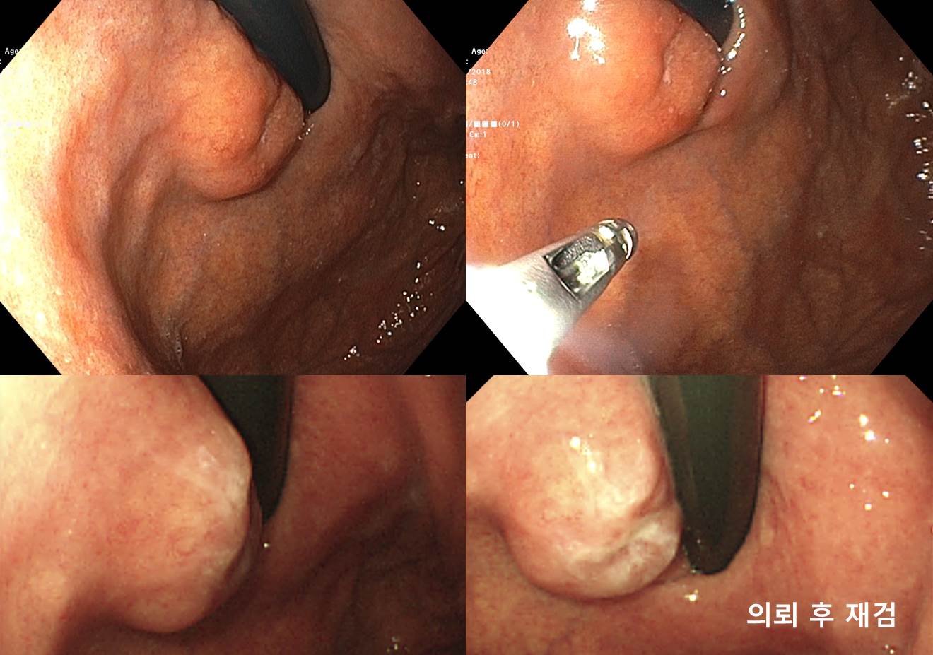
60세 여성 환자이십니다. 외과에서 ESD를 위하여 의뢰되셨습니다. 병소는 cardia에 닿아 있는 약 2cm 크기의 융기형 병소였고 그 중앙이 함몰되어 있었습니다. 함몰부를 제외한 융기부의 표면 점막은 정상으로 관찰되었습니다. 외부 조직검사 슬라이드 재판독은 moderately differentiated tubular adenocarcinoma였습니다. 의뢰 후 재검을 하였을 때 동일 소견이 보였는데 다만 전 내시경 시 조직검사 때문인지 함몰부가 다소 불규칙한 모습을 보이고 있었습니다. 당연히 수술이 필요한 상황인데 환자분께서 내시경 치료를 원하셨다고 합니다. 어떻게 하시겠습니까?
병소의 위치관계로 total gastrectomy, 최소한 proximal gastrectomy가 필요한 상황입니다. 물론 이와 같은 큰 수술을 받고 싶은 환자는 없을 것입니다. 수술 대신 내시경으로 치료할 수 있다면 내시경 치료를 시도하는 것이 당연한 원칙이겠지요. 그러나 작지만 뚜렷한 mass effect를 가지고 병소이고 중앙에 명백한 함몰형 병소가 있으므로 deep SM invasion을 의심할 수 있으므로 내시경 치료는 되지 않는 병소입니다. 환자분께서는 섭섭해 하셨을 수도 있겠지만 원칙에 따라 다소 냉정하게 수술적 치료를 권하였습니다. 환자에게 끌려서 부적당한 치료를 제공하는 것은 옳지 않은 일이라고 생각합니다.
최종 병리결과입니다. Gastritis cystica profunda를 동반한 deep SM cancer로 나왔습니다.
Stomach, total gastrectomy: Early gastric carcinoma
1. Location : upper third, Center at cardia (Siewert II) and posterior wall
2. Gross type : EGC type IIa
3. Histologic type : tubular adenocarcinoma, moderately differentiated
4. Histologic type by Lauren : intestinal
5. Size : 1.7x1.2 cm
6. Depth of invasion : invades submucosa (sm3) (pT1b)
7. Resection margin: free from carcinoma, safety margin: proximal 1 cm, distal 15 cm
8. Lymph node metastasis : no metastasis in 27 regional lymph nodes (pN0) (0/27: "2", 0/1; "3", 0/4; "4", 0/3; "5", 0/1; "6", 0/4; "7", 0/3; "9", 0/1; "8a", 0/4; "11p", 0/3; "12a", 0/0; "4sb", 0/0; "1", 0/3)
9. Lymphatic invasion : not identified
10. Venous invasion : not identified
11. Perineural invasion : not identified
12. Associated findings : gastritis cystica profunda
13. AJCC stage by 8th edition: pT1b N0
현미경 소견입니다.


Deep SM layer
Mucosal surface
© 일원내시경교실 바른내시경연구소 이준행. EndoTODAY Endoscopy Learning Center. Lee Jun Haeng. (2019-2-26, update 2024-12-9)