EndoTODAY 내시경 교실
Beginner | ESA | Schedule | OPD
Seminars | Atlas | Recent | Links
 [Gastric cancer 604]
001 | 101 | 201 | 301 | 401 | 501 | 601 | 701 | 801 | 901 | 1000
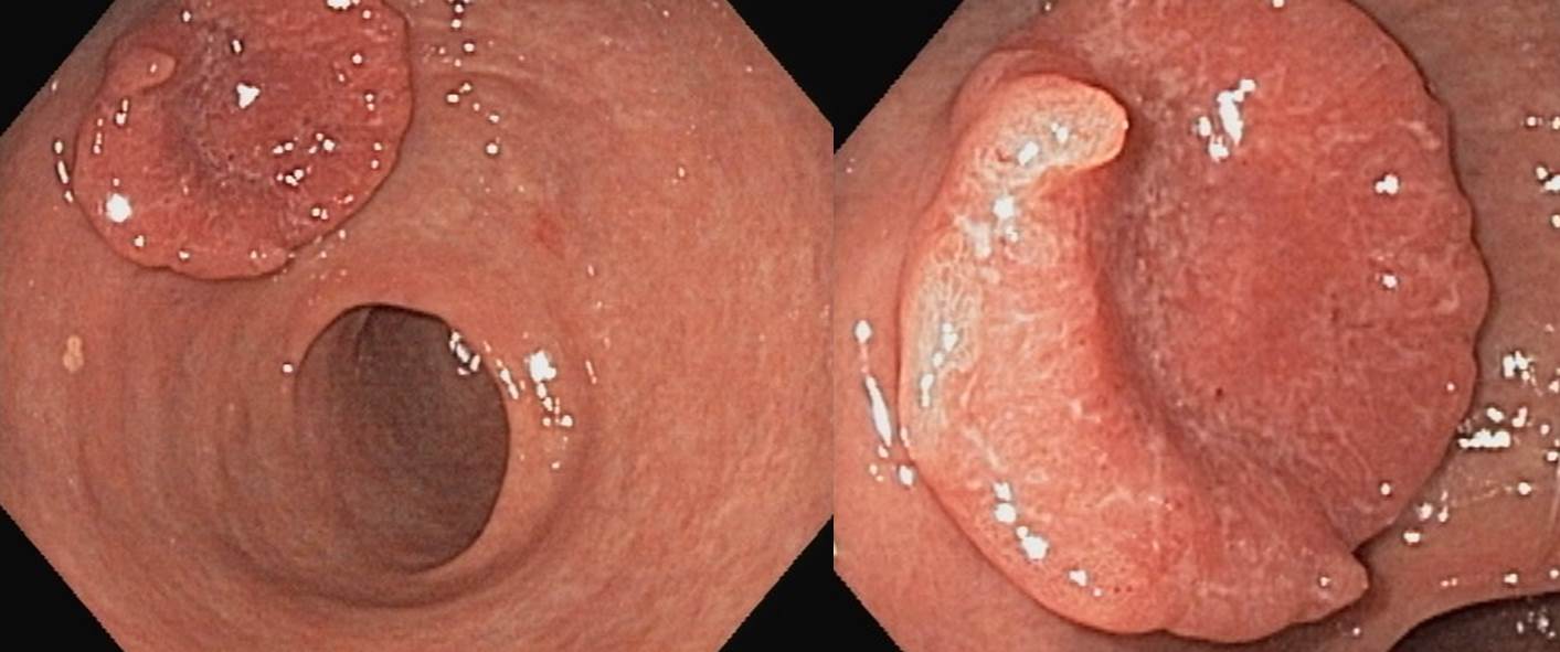
검진 내시경에서 발견된 AGC입니다. Mass의 일부가 함몰되었지만 그래도 전체적으로는 융기된 모양으로 AGC Borrmann type I으로 진단하였습니다. 조직검사는 tubular adenocarcinoma, moderately differentiated with neuroendocrine differentiation으로 보고되었습니다.
Stomach, subtotal gastrectomy: Advanced gastric carcinoma
1. Location : lower third, Center at antrum and lesser curvature
2. Gross type : Borrmann type 2
3. Histologic type : tubular adenocarcinoma, moderately differentiated with focal neuroendocrine differentiation
4. Histologic type by Lauren : intestinal
5. Size : 3.5x4.5 cm
6. Depth of invasion : penetrates subserosal connective tissue (pT3)
7. Resection margin: free from carcinoma, safety margin: proximal 4.5 cm, distal 4 cm
8. Lymph node metastasis : metastasis to 5 out of 52 regional lymph nodes (pN2) (perinodal extension: present) (5/52: "3", 2/17; "4", 1/14; "5", 1/2; "6", 0/4; "7", 0/2; "8a", 0/2; "9", 0/1; "11p", 0/2; "12a", 1/3; "4sb", 0/4; "1", 0/0; "4d", 0/1)
9. Lymphatic invasion : not identified
10. Venous invasion : not identified
11. Perineural invasion : not identified
12. Associated findings : ulceration
13. Peritoneal cytology : negative
14. AJCC stage by 7th edition: pT3 N2
증례와 관련하여 3가지 정도의 생각해 보았습니다.
+++ 1 +++
내시경 의사는 보만 1형으로 주었는데 병리의사는 보만 2형으로 주었습니다. 흔히 있는 일입니다. 약간 궤양이 있는 것은 사실이니까... 보통 비교적 둥그렇고 궤양이 깊고 궤양 주변의 dam이 두껍지 않고 현저히 융기되어 있으면 보만 2형이라고 부르고 있습니다. 그냥 덩어리이고 중앙이 약간 함몰되어 있으면 보만 1형으로 부릅니다. 주관적입니다.
+++ 2 +++
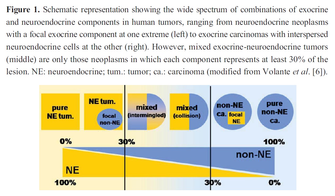
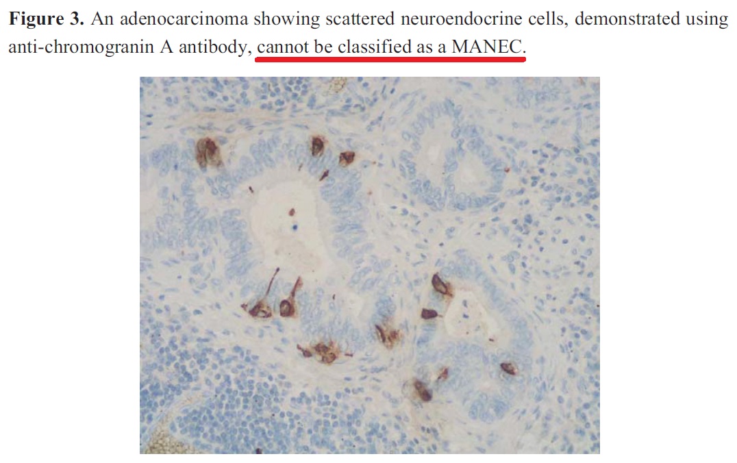
Forceps biopsy와 surgical pathology에서 모두 focal neuroendocrine differentiation의 언급이 있습니다. 일반적인 adenocarcinoma (이는 exocrine tumor라고 할 수 있습니다. Gastric epithelium에서 발생하는 tumor이기 때문입니다)에 약간의 endocrine component가 동반된 경우는 간혹 있습니다. 치료는 똑같습니다.
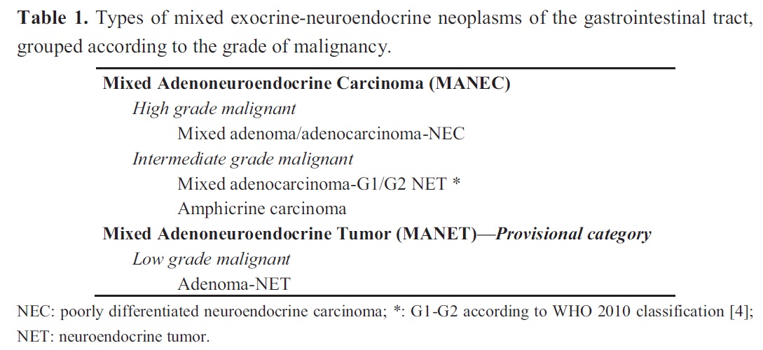
그러나 exocrine component와 endoscrine component가 대등하면 amphicrine tumor라고 합니다. 기준은 상당히 작위적이지만 대강 30%로 잡고 있습니다. 최근에는 Mixed Adenoneuroendocrine Carcinomas (MANECs)라는 이름으로 통일되고 있는 것 같습니다 (Cancers 2012;4:11-30).
Amphicrine tumor의 특징은 아래과 같습니다.
+++ 3 +++
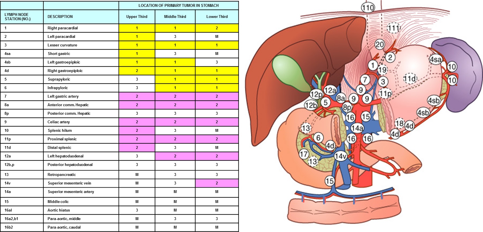
12a 림프절 전이가 있었습니다. Left hepatoduodenal node입니다. 이는 과거 방식으로는 group 2 station입니다. 위암이 mid 1/3 혹은 distal 1/3에서는 group 2로 proximal 1/3에서는 group 3로 불렀습니다.
© 일원내시경교실 바른내시경연구소 이준행. EndoTODAY Endoscopy Learning Center. Lee Jun Haeng.