 EndoTODAY 내시경 교실
Beginner | ESA | Schedule | OPD
Seminars | Atlas | Recent | Links
 [Gastric cancer 519 - ESD 환자에서 우연히 발견된 담낭암]
001 | 101 | 201 | 301 | 401 | 501 | 601 | 701 | 801 | 901 | 1000
위암 ESD 환자의 시술 전 CT에서 위 이외의 장기에서 incidental finding이 발견되곤 합니다. 이번 증례는 담낭암이 우연히 발견된 경우입니다. 위 ESD 후 담낭 수술을 하였습니다.
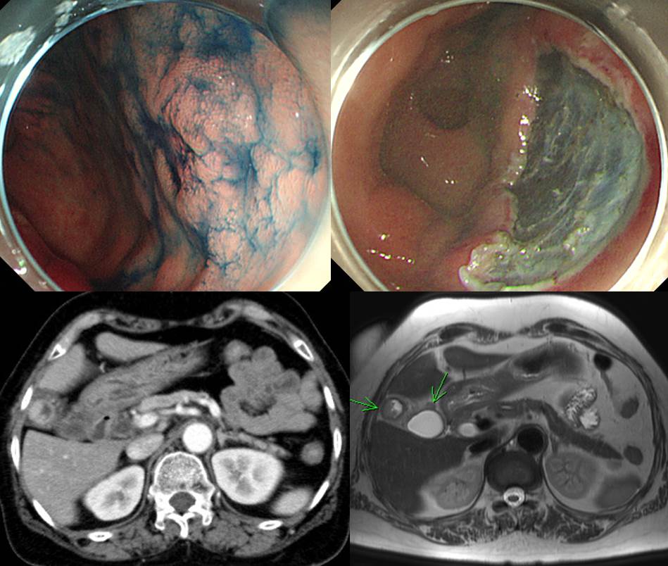
Posterior wall of antrum, endoscopic submucosal dissection:
. Early gastric carcinoma
1. Location : antrum, posterior wall
2. Gross type : EGC type IIc
3. Histologic type : tubular adenocarcinoma, moderately differentiated
4. Histologic type by Lauren : intestinal
5. Size of carcinoma : (1) longest diameter, 5 mm (2) vertical diameter, 4 mm
6. Depth of invasion : invades mucosa (lamina propria) (pT1a)
7. Resection margin : free from carcinoma(N), safety margin : distal 10 mm, proximal 5 mm, anterior 4 mm, posterior 12 mm
8. Lymphatic invasion : not identified(N)
9. Venous invasion : not identified(N)
10. Perineural invasion : not identified(N)
11. Microscopic ulcer : absent
12. Histologic heterogeneity: absentGALLBLADDER CANCER arising from flat dysplasia
III. Histopathologic Diagnosis
(1) Histologic type: Adenocarcinoma
(2) Histologic grade: G2 (moderately differentiated)
(3) Invasive carcinoma size: greatest dimension (3cm), depth of invasion (0.7cm)
(4) Precursor lesion: flat dysplasia
(5) T2: Tumor invades perimuscular connective tissue; no extension beyond serosa or into liver
(6) Involvement of large vessel: absent
(7) N0: No regional lymph node metastases (0/15: "LN7,8,9", 0/6; "LN12", 0/4; "LN13", 0/4; pericholedochal, 0/1)
(8) M0: No distant metastasis
(9) Margin status
- Cystic duct margin: negative (negative margin: 1cm)
- Liver capsule margin: negative (safety margin: 1cm)
(10) Perineural and neural invasion: present
(11) Lymphovascular invasion: present
(12) Involvement of Rokitansky-Ascoff Sinus by high-grade BilIN: present[Additional note]
(1) Proportion: well to moderately (100%) / poorly (0%) differentiated
(2) Pattern Score (PS): 4(2+2)
(3) Location: fundus and body
(4) Mucoepidermoid-like feature: absent
(5) Stromal reaction: desmoplasia (1) / inflammation (1)
(6) Tumor necrosis: absent
(7) Nuclear pleomorphism: moderate
(8) Mucin production: abortive
(9) Tumor Regression grade: Not applicable
(10) ICD code: M8500GBca
R0: complete resection with grossly and microscopically negative resection margins
AJCC Stage Groupings (2010, 7th Ed)
Stage II T2 N0 M0
일전에 비슷한 증례를 소개드린 바 있습니다 (EndoTODAY 위암 318). 당시에는 ESD 후 추적관찰 CT에서 담낭암이 우연히 발견되었던 경우였습니다. 아래에 옮깁니다.
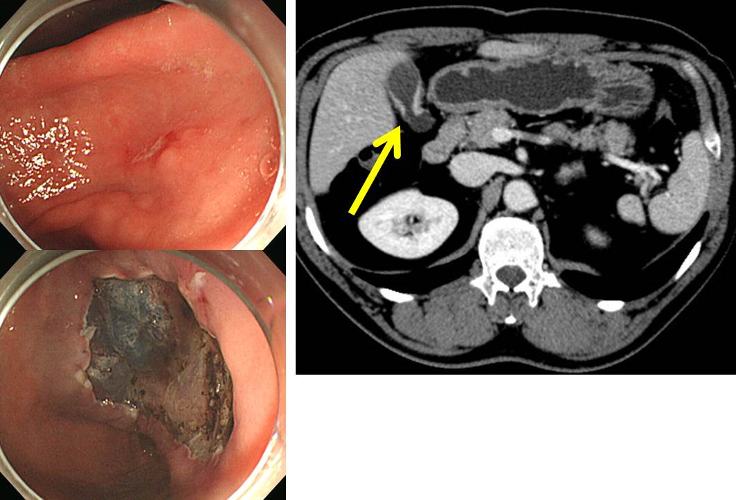
Stomach, endoscopic submucosal dissection:
. Early gastric carcinoma
1. Location : antrum, lesser curvature
2. Gross type : EGC type IIb
3. Histologic type : tubular adenocarcinoma, moderately differentiated
4. Histologic type by Lauren : intestinal
5. Size of carcinoma : (1) longest diameter, 20 mm (2) vertical diameter, 12 mm
6. Depth of invasion : invades mucosa (lamina propria) (pT1a)
7. Resection margin : free from carcinoma(N), safety margin : distal 6 mm, proximal 5 mm, anterior 8 mm, posterior 12 mm
8. Lymphatic invasion : not identified(N)
9. Venous invasion : not identified(N)
10. Perineural invasion : not identified(N)
11. Microscopic ulcer : absent
12. Histologic heterogeneity: absentGALLBLADDER CANCER arising from flat dysplasia
(1) Histologic type: Adenocarcinoma
(2) Histologic grade: G3 (poorly differentiated)
(3) Invasive carcinoma size: greatest dimension (2.8cm), depth of invasion (0.8cm)
(4) Precursor lesion: flat dysplasia
(5) T2: Tumor invades perimuscular connective tissue; no extension beyond serosa or into liver
(6) Involvement of large vessel: absent
(7) N0: No regional lymph node metastases (0/2: "LN12", 0/0; "LN12 & 8", 0/2)
(8) M0: No distant metastasis
(9) Margin status
- Cystic duct margin: negative (safety margin: 2.1 cm)
- Liver deep (radial) margin: negative
(10) Perineural and neural invasion: present
(11) Lymphovascular invasion: present
(12) Involvement of Rokitansky-Ascoff Sinus by high-grade BilIN: not applicable
(13) No evidence of malignancy and focal eosinophilic abscess (0.3 cm), "liver nodule" for frozen section-3[Additional note]
(1) Proportion: well to moderately (10 %) / poorly (90 %) differentiated
(2) Pattern Score (PS): 6 (3+3)
(3) Location: fundus
(4) Mucoepidermoid-like feature: absent
(5) Stromal reaction: desmoplasia (2) / inflammation (0) / myxoid change (2)
(6) Tumor necrosis: absent
(7) Nuclear pleomorphism: moderate
(8) Mucin production: abortive
(9) Tumor Regression grade: Not applicable
(10) ICD code: M8500GBca
R0: complete resection with grossly and microscopically negative resection margins
AJCC Stage Groupings (2010, 7th Ed)
Stage II T2 N0 M0
© 일원내시경교실 바른내시경연구소 이준행. EndoTODAY Endoscopy Learning Center. Lee Jun Haeng. (2017-9-14)