EndoTODAY 내시경 교실
Beginner | ESA | Schedule | OPD
Seminars | Atlas | Recent | Links
 [Gastric cancer 499]
001 | 101 | 201 | 301 | 401 | 501 | 601 | 701 | 801 | 901 | 1000
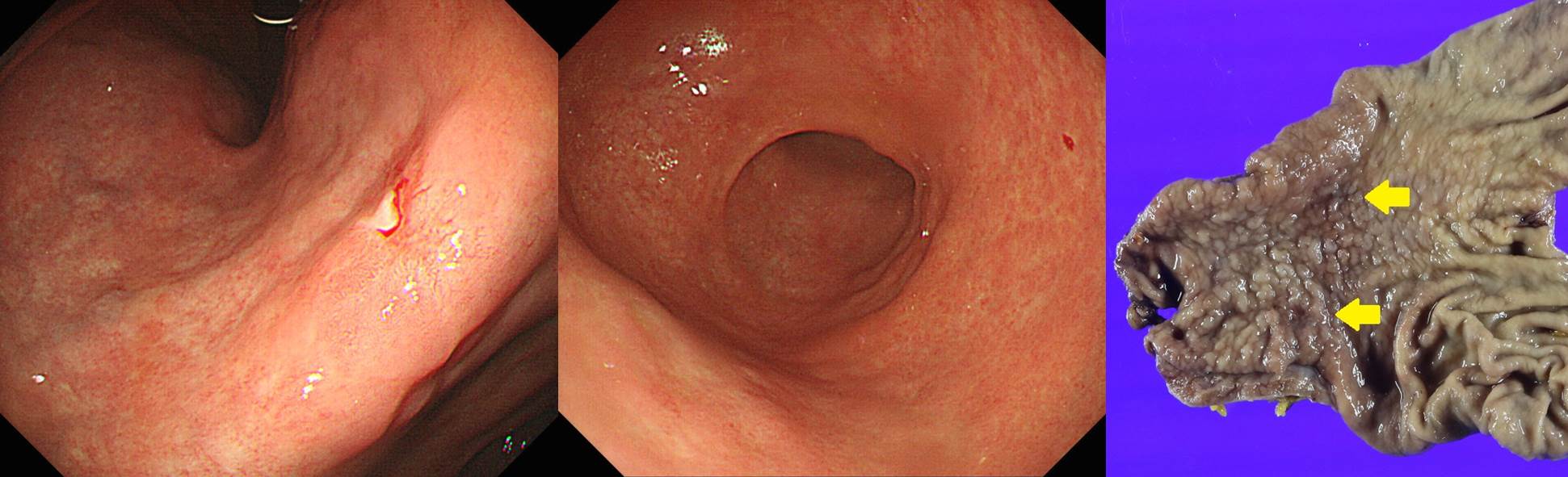
수술 전 내시경 검사에서 암이 하나였는데, 수술 후 최종 병리에서 암이 두개로 보고되는 예가 있습니다. 첫 내시경 사진을 잘 살펴보아도 두번째 병소를 찾지 못하는 경우가 대부분입니다.
최종 병리 결과를 옮깁니다.
1-3. Stomach, subtotal gastrectomy:
. Multiple gastric carcinomas (x2)
I. Early gastric carcinoma
1. Location : lower third, Center at angle and lesser curvature
2. Gross type : EGC type IIc
3. Histologic type : signet-ring cell carcinoma
4. Histologic type by Lauren : mixed
5. Size : 2x1 cm
6. Depth of invasion : invades mucosa (muscularis mucosae) (pT1a)
7. Resection margin: free from carcinoma, safety margin: proximal 2.3 cm, distal 7.2 cm
8. Lymphatic invasion : not identified
9. Venous invasion : not identified
10. Perineural invasion : not identified
II. Early gastric carcinoma
1. Location : middle third, Center at angle and anterior wall
2. Gross type : EGC type IIc
3. Histologic type : signet-ring cell carcinoma
4. Histologic type by Lauren : diffuse
5. Size : 1x1 cm
6. Depth of invasion : invades mucosa (lamina propria) (pT1a)
7. Resection margin: free from carcinoma, safety margin: proximal 2.5 cm, distal 7.2 cm
8. Lymphatic invasion : not identified
9. Venous invasion : not identified
10. Perineural invasion : not identified
III.
1. Lymph node metastasis : no metastasis in 55 regional lymph nodes (pN0) (0/55: "3", 0/16; "4", 0/16; "5", 0/1; "6", 0/5; "7", 0/3; "9", 0/2; "8a", 0/7; "11p", 0/1; "12a", 0/2; "4sb", 0/0; "1", 0/2)
2. AJCC stage by 7th edition: pT1a N0
. No evidence of malignancy, "EEA ring"
(본 진단은 조직구축학적 검사 후 판독결과 입니다.)
<< Addendum>>
Specimen condition: paraffin block
<< Results of immunohistochemistry>>
. c-erbB-2(HER2): Negative (0)
Note: We use FDA-approved Ventana PATHWAY anti-HER-2/neu (4B5) Rabbit Monoclonal Antibody for this assay.
*** Staining for positive and negative control, and negative reagent control was performed. All controls show appropriate reactivity.
<< Addendum>>
Specimen condition : paraffin block
<< Result of immunohistochemistry>>
. Cytokeratin AE1/AE3 PAN CK : Negative
<< Addendum>>
Specimen condition: paraffin block
<< Result of in-situ hybridization>>
. Epstein-Barr virus: Negative (CA, conventional adenocarcinoma)
*** Staining for positive and negative control, and negative reagent control was performed. All controls show appropriate reactivity.
【GROSS DESCRIPTION 】
1. FS-1
Received fresh labeled "proximal margin" is a mucosal strip, measuring 1cm.
Entirely embedded.
2. FS-2
Received fresh labeled "distal margin" is a mucosal strip, measuring 1cm.
Entirely embedded.
3.
Received fresh is a subtotally resected stomach with the omentum in continuity. The stomach is previously opened along the greater curvature by the surgeon and measures 16.5 cm along the greater curvature and 10 cm along the lesser curvature. The serosal surface is smooth and glistening. The mucosal surface shows a depressed lesion (EGC type IIc), measuring 1x0.6 cm, at the lesser curvature of body. The lesion is located 2.2 cm apart from the proximal resection margin and 6.5 cm apart from the distal resection margin. All perigastric lymph nodes found are embedded.
REPRESENTATIVE SECTIONS.
3A: "3"
3B: "4"
3C: "5"
3D: "6"
3E: "7"
3F: "9"
3G: "8a"
3H: "11p"
3I: "12a"
3J: "4sb"
3K: "1"
3L-N: MAPPING
Frozen section diagnosis
FS-1: "proximal margin" - No tumor (JY)
FS-2: "distal margin" - No tumor (JY)
[Block Information]
1 proximal margin
2 distal margin
3A lymph node #3
3B lymph node #4
3C lymph node #5
3D lymph node #6
3E lymph node #7
3F lymph node #9
3G lymph node #8a
3H lymph node #11p
3I lymph node #12a
3J lymph node #4sb
3K Lymph node #1
3L ~ N Stomach
© 일원내시경교실 바른내시경연구소 이준행. EndoTODAY Endoscopy Learning Center. Lee Jun Haeng.