 EndoTODAY 내시경 교실
Beginner | ESA | Schedule | OPD
Seminars | Atlas | Recent | Links
 [Gastric cancer 444]
001 | 101 | 201 | 301 | 401 | 501 | 601 | 701 | 801 | 901 | 1000
조기위암 내시경 치료 후 경과관찰 도중 온갖 암들이 발견됩니다. 이번에는 전립선암이었습니다.
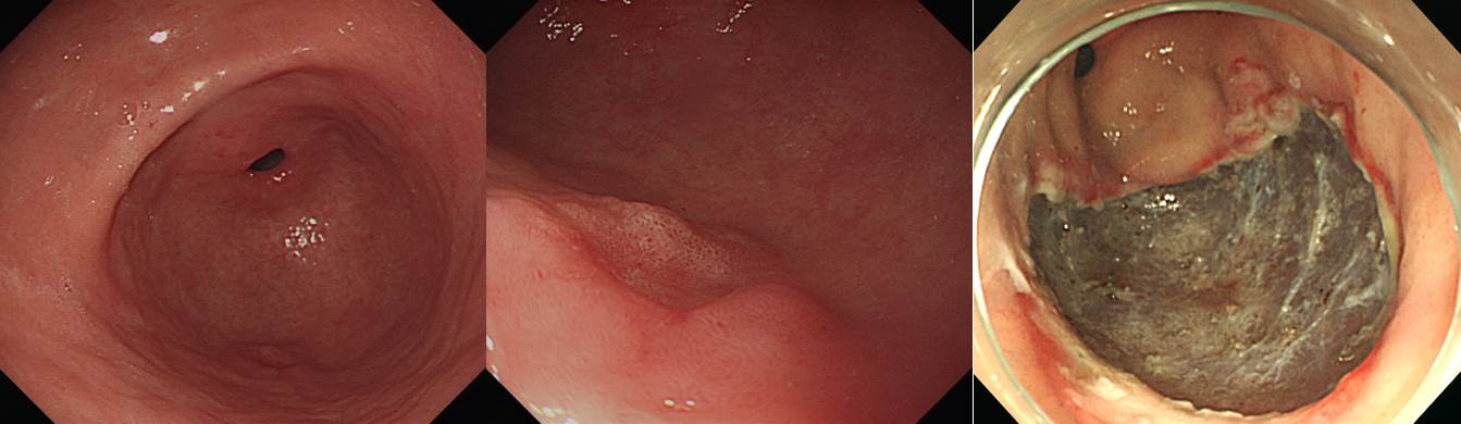
Stomach, endoscopic submucosal dissection:
Early gastric carcinoma
1. Location : mid antrum, greater curvature
2. Gross type : EGC type IIc
3. Histologic type : tubular adenocarcinoma, moderately differentiated (90%) >> poorly differentiated (10%)
4. Histologic type by Lauren : intestinal
5. Size of carcinoma : (1) longest diameter, 14mm (2) vertical diameter, 8mm
6. Depth of invasion : invades mucosa (muscularis mucosa) (pT1a)
7. Resection margin : free from carcinoma(N), safety margin : distal 16 mm, proximal 9 mm, anterior 10 mm, posterior 14 mm
8. Lymphatic invasion : not identified(N)
9. Venous invasion : not identified(N)
10. Perineural invasion : not identified(N)
11. Microscopic ulcer : absent
12. Histologic heterogeneity: present
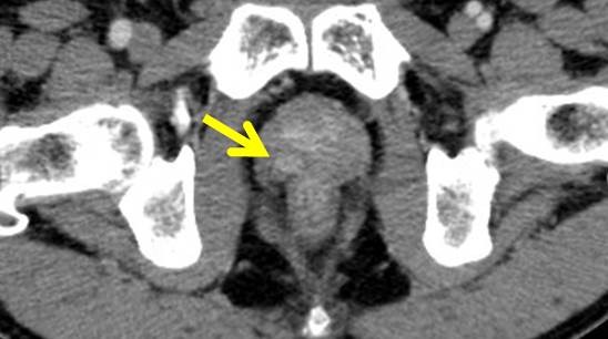
Radical prostatectomy:
Adenocarcinoma(s) (x2), total tumor volume about 10%
1) tumor(s): (a) 1.9x0.9x2 cm, Gleason's score 3+4=7/10 (peripheral zone of right lobe), (b) 0.7x0.3x0.4 cm, Gleason's score 3+4=7/10 (peripheral zone of left lobe)
2) no extraprostatic extension
3) no involvement of bilateral seminal vesicles and negative vas
4) resection margin: apex (negative), base (negative), body (negative)
5) no involvement of bilateral apex
6) no involvement of bilateral base
7) perineural invasion: present
8) lymphovascular invasion: not identified
9) high grade prostatic intraepithelial neoplasia : present
10) AJCC pathologic stage pT2c R0
© 일원내시경교실 바른내시경연구소 이준행. EndoTODAY Endoscopy Learning Center. Lee Jun Haeng.