EndoTODAY 내시경 교실
Beginner | ESA | Schedule | OPD
Seminars | Atlas | Recent | Links
 [Gastric cancer 438]
001 | 101 | 201 | 301 | 401 | 501 | 601 | 701 | 801 | 901 | 1000
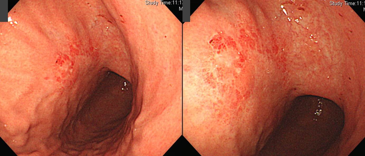
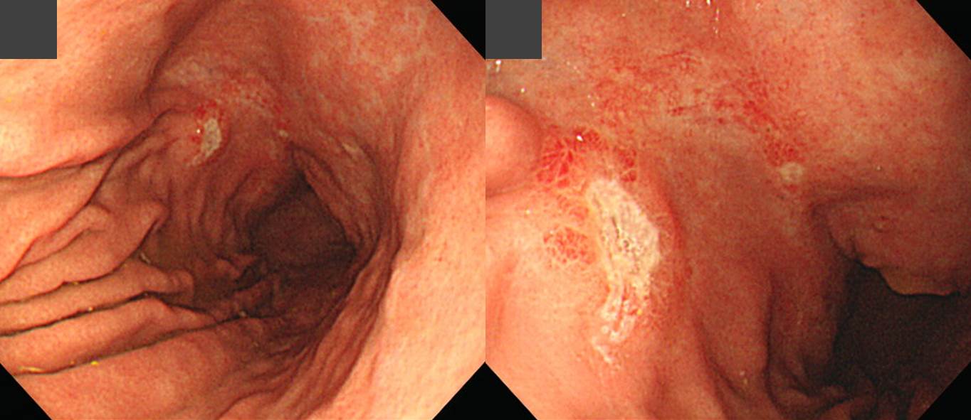
조직검사 후 간혹 출혈이 발생할 수 있는데 위암의 경우도 예외는 아닙니다.
"위체하부 전벽에 약 4cm 크기의 경계가 불분명하고 표면이 불규칙한 발적과 퇴색이 혼재한 영역이 있음. 여러 방향에서 점막주름이 끌려오고 있으며 병소의 경계 부근에서 끊김." 정도의 소견으로 EGC IIb 임상진단 하에 조직검사를 하였습니다. Melena가 있어서 내시경 재검을 하였습니다. 조직검사로 인한 뚜렷한 궤양이 관찰되었습니다.
수술을 하였고 추정하였던 것보다 큰 것으로 나왔습니다. Signet ring cell carcinoma에서 흔히 있는 일입니다.
Early gastric carcinoma
1. Location : middle third, center at body and anterior wall
2. Gross type : EGC type IIb
3. Histologic type : signet-ring cell carcinoma
4. Histologic type by Lauren : indeterminate
5. Size : 6.5x3.3 cm
6. Depth of invasion : extension to mucosa (muscularis mucosa) (pT1a)
7. Resection margin: free from carcinoma, safety margin: distal 5.2 cm, proximal 4 cm
8. Lymph node metastasis : no metastasis in 33 regional lymph nodes (pN0)
9. Lymphatic invasion : not identified
10. Venous invasion : not identified
11. Perineural invasion : not identified
* 참고: EndoTODAY 위내시경 조직검사 후 출혈
© 일원내시경교실 바른내시경연구소 이준행. EndoTODAY Endoscopy Learning Center. Lee Jun Haeng.