 EndoTODAY 내시경 교실
Beginner | ESA | Schedule | OPD
Seminars | Atlas | Recent | Links
 [Gastric cancer 429]
001 | 101 | 201 | 301 | 401 | 501 | 601 | 701 | 801 | 901 | 1000
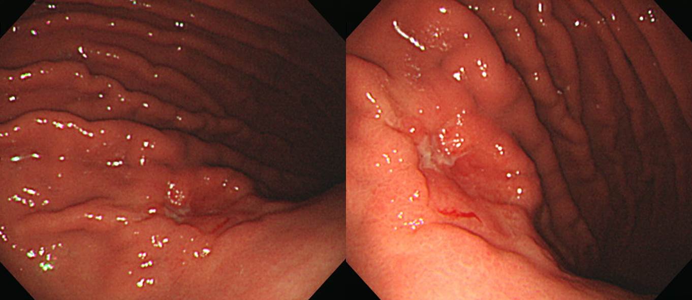
Stomach, total gastrectomy:
Advanced gastric carcinoma
1. Location : middle third, Center at body and posterior wall
2. Gross type : Borrmann type 3
3. Histologic type : signet-ring cell carcinoma
4. Histologic type by Lauren : diffuse
5. Size : 3.5x3.2 cm
6. Depth of invasion : penetrates serosa (pT3)
7. Resection margin: free from carcinoma, safety margin: proximal, 3.5 cm; distal, 14 cm
8. Lymph node metastasis : metastasis to 33 out of 48 regional lymph nodes (pN3) (33/48: lesser curvature, 16/18; greater curvature, 3/3; "1", 0/1; "2", 4/6; "4sb", 3/3; "5", 0/0; "6", 1/2; "8a", 0/3; "7", 3/3; "9", 0/2; "11p", 3/3; "12a", 0/4; "14v", 0/0)
9. Lymphatic invasion : present
10.Venous invasion : not identified
11.Perineural invasion : present
수술과 항암치료 후 5년 이상 NED (no evidence of disease) 상태로 지내다가 변비로 대장내시경을 하였습니다. 병소의 위치는 T colon으로 판단되는 AV 60 cm 부터 55cm까지의 diffuse mucosal edema, friability, luminal narrowing 소견이었습니다.
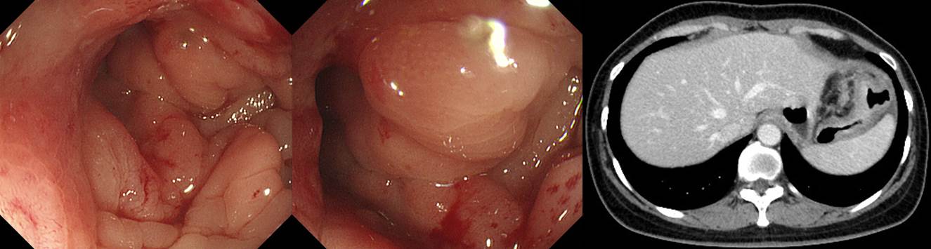
Biopsy: poorly differentiated carcinoma, favor metastatic
CT: Distal T-colon를 따라 enhancing wall thickening 및 주위 enlarged lymph node들이 관찰되는데 Bx. proven metastatic lesion으로 판단됨.
보만 4형 진행성 위암이 복막전이를 보이면 rectal obstruction을 일으키지만, 위암의 국소 재발에서는 T colon obstruction이 많습니다.
© 일원내시경교실 바른내시경연구소 이준행. EndoTODAY Endoscopy Learning Center. Lee Jun Haeng.