 EndoTODAY 내시경 교실
Beginner | ESA | Schedule | OPD
Seminars | Atlas | Recent | Links
 [Gastric cancer 403]
001 | 101 | 201 | 301 | 401 | 501 | 601 | 701 | 801 | 901 | 1000
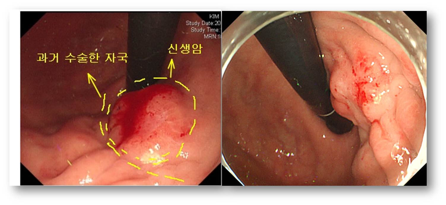
10년 전 위암 수술 후 잔위에서 발생한 위암입니다. 과거 suture line에 가까워 모양을 정확히 파악하기는 어려웠습니다. 수술을 시행하였고 생각보다 깊어서 놀랐습니다.
Status post subtotal gastrectomy due to advanced gastric carcinoma (S07-23212)
Advanced gastric carcinoma
1. Location : upper third, Center at cardia (Siewert III) and lesser curvature
2. Gross type : Borrmann type 3
3. Histologic type: tubular adenocarcinoma, poorly(poorly cohesive) differentiated
4. Histologic type by Lauren : diffuse
5. Size : 3.5x3 cm
6. Depth of invasion : invades adjacent liver parenchyme (pT4b)
7. Resection margin: free from carcinoma
8. Lymph node metastasis : no metastasis in 7 regional lymph nodes (pN0)
9. Lymphatic invasion : present
10. Venous invasion : not identified
11. Perineural invasion : present
12. AJCC stage by 7th edition: T4b N0
Poorly differentiated adenocarcinoma에서는 어지간해서는 수술하는 것이 좋겠습니다.
© EndoTODAY Endoscopy Learning Center. Jun Haeng Lee. 일원내시경교실 바른내시경연구소 이준행