 EndoTODAY 내시경 교실
Beginner | ESA | Schedule | OPD
Seminars | Atlas | Recent | Links
 [위암 319 - EndoTODAY 퀴즈 09]
001 | 101 | 201 | 301 | 401 | 501 | 601 | 701 | 801 | 901 | 1000
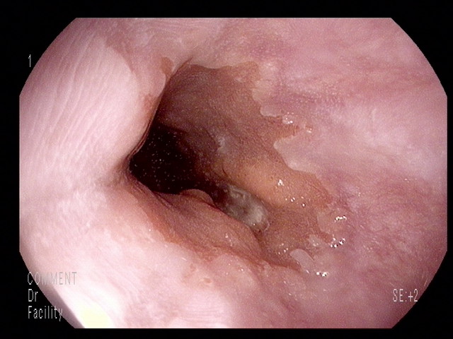
외부 사진 딱 한장으로 진단 맞춰보기 연습합시다. 첨부 사진을 보고 소견과 impression을 제게 메일로 보내주시기 바랍니다.
[애독자 답변]
답변 1: GEJ stomach side에 계곡처럼 움패인 ulcer와 margin은 elevation 된 소견이 보여서 AGC III 로 판단됩니다. → Impression: Cardia cancer
답변 2: 5mm 미만의 mucosal break가 보이고 치유되면서 백색혼탁이 약간씩 관찰되고 있습니다. → Impression: Reflux esophagitis LA-A
답변 3: Reflux esophagitis, Barrett esophagus with esophageal ulcer, R/O esaophageal cancer
답변 4: On the GE junction, irregular Z line was seen. On the gastric side, 0.6cm sized flat elevated lesion with nodularity was seen at the 7 o'clock direction. → Impression: Cardia adenoma, r/o cancer
답변 5: 3시와 6시 가운데에 점막의 주변부가 융기되어 있으며 중심부가 depression된 것으로 생각되며, 중심부에 exudate가 일부 덮혀 있습니다. → Impression: GEJ cancer.
[정답 및 해설]
문제가 너무 어려웠던 모양입니다. 환자를 의뢰하고 의뢰받는 과정에서는 명확하지 않은 사진을 보고 임상적 판단을 해야 하는 경우가 적지 않습니다. 저도 검사를 잘 했다고 생각한 후 다른 의사에게 증례를 소개할 때 좋은 사진이 없어서 곤란한 적이 많습니다.
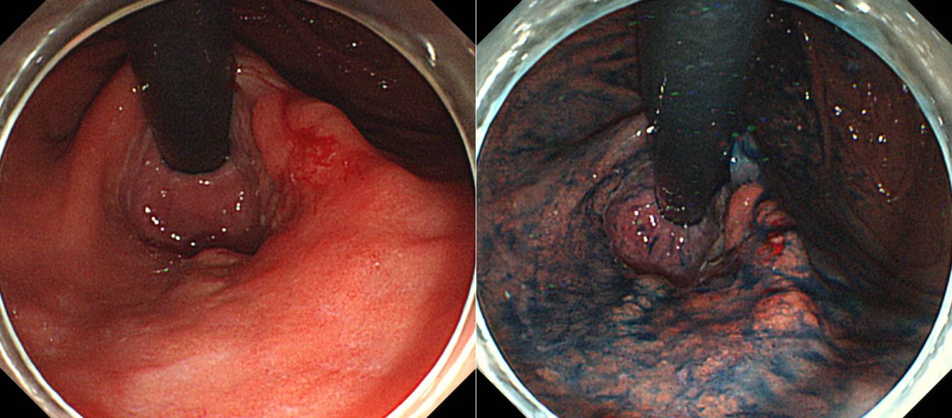
Cardia cancer였습니다. ESD 후 아쉽게도 deep SM invasion이 나왔습니다. 수술을 할 수 밖에 없습니다. 사실 이 부위 암은 생각보다 깊은 경우가 많습니다. 그렇다고 쉽사리 수술을 선택하기도 어렵습니다. Total gastrectomy가 될 것이므로....
Stomach, endoscopic submucosal dissection:
Early gastric carcinoma
1. Location : EG junction
2. Gross type : EGC type IIc
3. Histologic type : tubular adenocarcinoma, well differentiated
4. Histologic type by Lauren : intestinal
5. Size of carcinoma : (1) longest diameter, 28mm (2) vertical diameter, 18mm
6. Depth of invasion : invades submucosa, (depth of sm invasion : 750 ㎛) (pT1b)
7. Resection margin : free from carcinoma(N), safety margin : distal 1 mm, proximal 2 mm, anterior 6 mm, posterior 10 mm, deep 500㎛
8. Lymphatic invasion : not identified(N)
9. Venous invasion : not identified(N)
10. Perineural invasion : not identified(N)
11. Associated finding: gastritis cystica profunda
12. Microscopic ulcer : absent
13. Histologic heterogeneity: absent
© 2015-12-16. 일원내시경교실 바른내시경연구소 이준행