 EndoTODAY 내시경 교실
Beginner | ESA | Schedule | OPD
Seminars | Atlas | Recent | Links
 [위암 220]
001 | 101 | 201 | 301 | 401 | 501 | 601 | 701 | 801 | 901 | 1000
병변의 경계가 어디까지라고 생각되십니까?
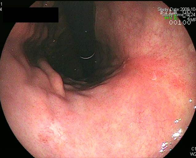
1. Location : middle third, center at body and lesser curvature
2. Gross type : EGC type IIc+III
3. Histologic type : tubular adenocarcinoma, poorly differentiated
4. Histologic type by Lauren : intestinal
5. Size : 5.2x4.3x0.1 cm
6. Depth of invasion : extension to mucosa (muscularis mucosa) (pT1a)
7. Resection margin: free from carcinoma, safety margin: proximal, 1.5 cm; distal, 5.1 cm
8. Lymph node metastasis : no metastasis in 43 regional lymph nodes (pN0)
9. Lymphatic invasion : not identified
10.Venous invasion : not identified
11.Perineural invasion : not identified
12.Associated findings : ulceration (ul III)
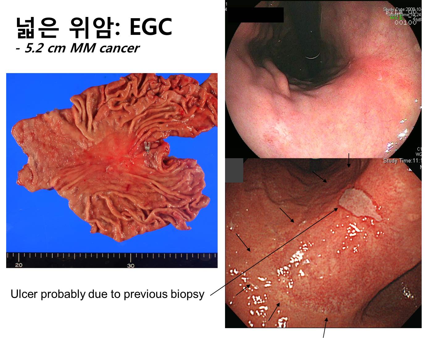
상당히 넓은 병소의 조기위암이라고 하더라도 점막에 국한된 경우는 얼마든지 있습니다. 우측 상단은 환자가 의뢰될 때 가져오신 사진이므로 조직 검사 전의 naive한 병소이고 우측 하단은 본 병원에서 재검한 사진이므로 조직검사 후의 모습입니다. 병소 중간의 ulcerative lesion은 조직검사에 의한 변화인 것으로 판단되었습니다.
이 환자에서 궤양에만 관심을 두고 그 주변을 예민하게 관찰하지 못한다면 비교적 큰 조기위암을 작은 것으로 오인할 수 있겠습니다. 수술 후 최종 크기는 5.2x4.3cm이었고 muscularis mucosa까지 가 있는 조기위암이었습니다. 우측 하단의 사진에서 제가 작은 화살표로 표시한 부분을 잘 관찰하시기 바랍니다. 암과 정상부위의 경계입니다.
 © 2015-3-16. 이준행