 EndoTODAY 내시경 교실
Beginner | ESA | Schedule | OPD
Seminars | Atlas | Recent | Links
 [위암 140]
001 | 101 | 201 | 301 | 401 | 501 | 601 | 701 | 801 | 901 | 1000
위식도접합부암이고 조직형이 poorly differentiated인 경우는 고민하지 않고 수술을 보냅니다. 생각보다 크거나 깊다고 나오는 경우가 종종 있습니다.
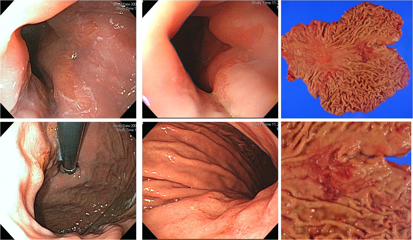
Early gastric carcinoma
1. Location : upper third, center at cardia and posterior wall
2. Gross type : EGC type IIc
3. Histologic type : tubular adenocarcinoma, poorly differentiated
4. Histologic type by Lauren : mixed
5. Size : 4.6x4 cm
6. Depth of invasion : extension to mucosa (muscularis mucosa) (pT1a)
7. Resection margin: free from carcinoma
8. Lymph node metastasis : no metastasis in 42 regional lymph nodes (pN0)
9. Lymphatic invasion : present
10. Venous invasion : not identified
11. Perineural invasion : not identified
 © 2014-11-25. 이준행