EndoTODAY ГЛНУАц БГНЧ
Beginner | ESA | Schedule | OPD
Seminars | Atlas | Recent | Links
![]() [Thursday Endoscopy Conference 20170810]
[Thursday Endoscopy Conference 20170810]
![]() 1. Rectal GIST
1. Rectal GIST
TEMРИЗЮ ФЁЗсЧЯПДДј rectal GISTРдДЯДй.
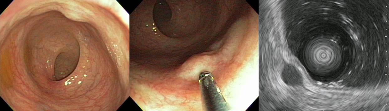
Rectum, trans-anal endoscopic microsurgery:
. Gastrointestinal stromal tumor of very low risk of malignant potential by proposed modification for adjuvant therapy (2008), none risk (0 %) of progressive disease by Miettinen (2006) :
1) tumor size: 1.4x1.2x0.4 cm
2) mitosis: 3/50 HPF (high powered fields)
3) histological type: spindle
4) necrosis: absent
5) cellularity: intermediate
6) cellular atypia: mild
7) invasion into mucosa: absent
8) resection margin involvement: absent
. Pho-H-H3: Positive in less than 5% of tumor cells
. C-kit : Positive
. DOG1 : Positive
. Ki-67: Negative
. PKC-ЅШ: Weakly positive
СІ РќАјРК ОЦДЯСіИИ... РЬЗИАд РлРК GISTДТ follow up ЧЯИщ ОюЖВАЁ Л§АЂЧи КИОвНРДЯДй.
![]() 2. Actinomycosis
2. Actinomycosis
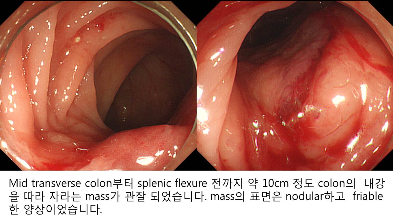
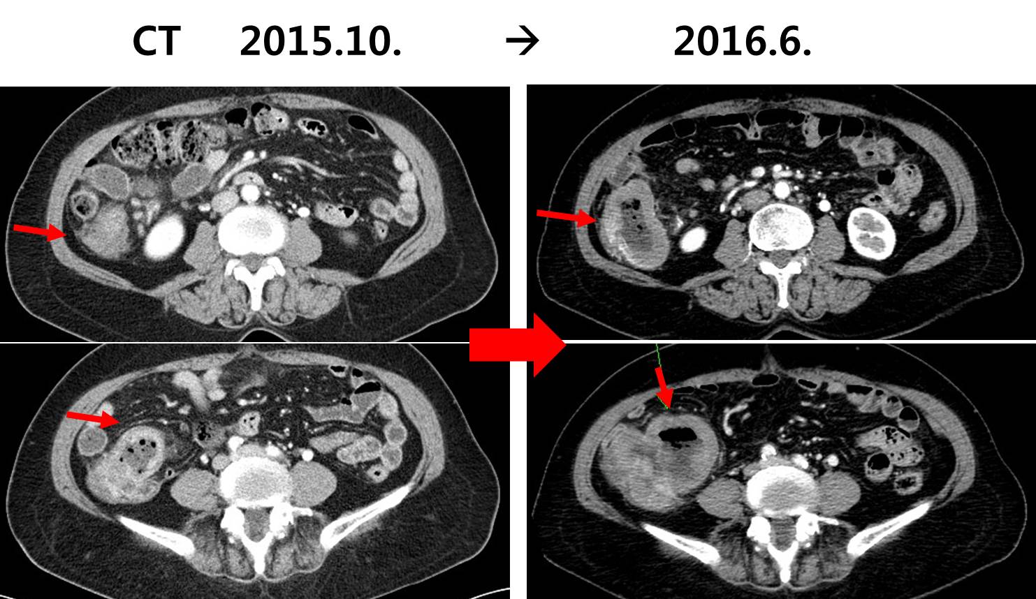
2014Гт СѕЗЪРдДЯДй. LUQ pain, hematochezia, weight loss 3~4kg/10days ЕюРИЗЮ CTИІ НУЧрЧЯАэ "colon(splenic flexure) wall thickening with omentum invasion РжОю r/o colon ca, r/o metastatic cancer" МвАпРИЗЮ РЧЗкЕЧОњНРДЯДй. ДыРхГЛНУАцАњ CT ЦЧЕЖРЛ ЧЯПДНРДЯДй. CT ЦЧЕЖРЬ ИХПь ШЧИЂЧпНРДЯДй.
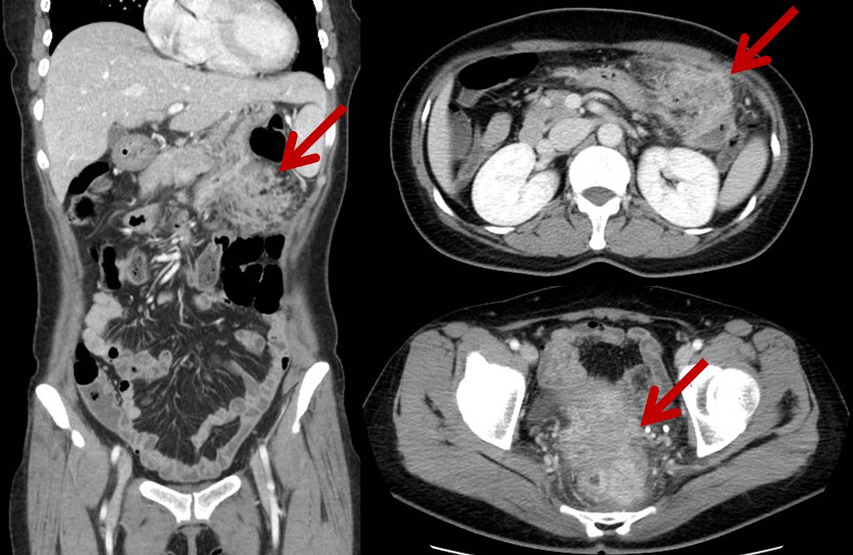
CT ЦЧЕЖ: Left sideРЧ gastrocolic ligamentИІ ЕћЖѓМ irregular enhancing massАЁ РжРН. АцАшАЁ СССі ОЪОЦ ХЉБтРЧ УјСЄПЁ СІЧбРЬ РжРИГЊ 6 cm РЬЛѓРЧ extentИІ КИРг. РЬ КДКЏРК ЛѓЙцРИЗЮ stomachРЧ body greater curvature sideПЭ ЧЯЙцРИЗЮДТ transverse colonРЧ upper wallБюСі extensionЧЯАэ РжРН. InvolvementЕШ bowel wall thickeningРЬ РжРИГЊ КёБГРћ layeringРЬ РЏСіЕЧАэ РжРН. БзЗЏГЊ transverse colonПЁМДТ focalЧЯАд layeringРЬ РЏСіЕЧСі ОЪДТ КЮКаРЬ РЧНЩЕЪ. Left side mesorectumПЁ Ор 3.3 cm sizeРЧ enhancing massАЁ РжАэ ПЊНУ АцАшАЁ СССі ОЪРИИч СжКЏРИЗЮ infiltrationРЛ ЕПЙнЧЯАэ РжРН. RectumРЧ left side wallАњ abuttingЧЯАэ РжРИИч РЮСЂЧб rectal wallПЁ wall thickeningРЬ РжАэ ПЊНУ layeringРЬ РЏСіЕЧАэ РжРН. Pelvic cavityПЁ МвЗЎРЧ fluid collectionРЬ РжРН. UterusПЁ IUD insertion stateРг. КЙА ГЛ РЧЙЬРжАд ФПСј lymph node КИРЬСі ОЪРН. Бз Пм liverПЭ spleen, pancreas, both kidneysПЁ ЦЏРЬМвАп ОјРН. GBПЁ ЦЏРЬМвАп ОјАэ biliary tree dilatation ОјРН. ScanПЁ ЦїЧдЕШ basal lungАњ boneПЁ ЦЏРЬМвАп ОјРН.
CONCLUSION: Inflammatory lesion such as actinomycosis R/O Malignancy such as transverse colon cancer with peritoneal seeding.
RECOMMENDATION: Transrectal biopsy for mesorectal mass.
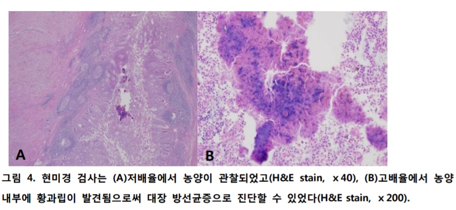
Transvaginal biopsyИІ НУЧрЧЯПДАэ ДйРНРЧ АсАњПДНРДЯДй. Inflamed granulation tissue with abscess and dense fibrosis. Bacterial colony present, consistent with actinomycosis
Treatment: IV penicillin G
2017Гт 3Пљ ГЛНУАцЧаШИ БГРАРкЗсАЁ КЙКЮ ЙцМББеСѕ (actinomycosis) РЬОњНРДЯДй.
![]() 3. Colon lymphoma
3. Colon lymphoma
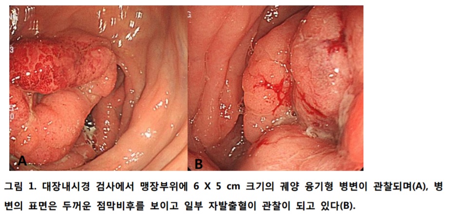
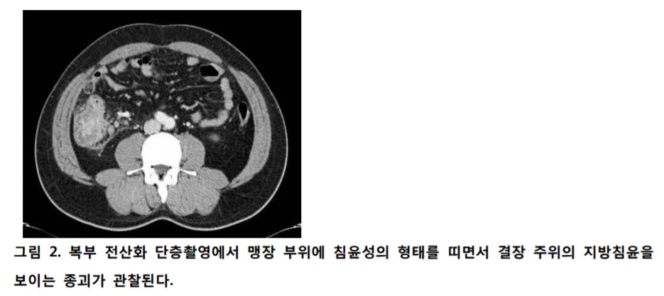
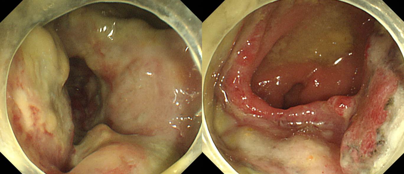
CecumАњ proximal A colonРЧ diverticulitis МвАпРЬГЊ СѕЛѓ, РЇФЁАЁ correlation ЕЧСі ОЪОЦ further evaluation РЇЧЯПЉ АцАњАќТћ Сп РЧЗкЕШ КаРдДЯДй.
СяНУ ДыРхГЛНУАцРЛ ДйНУ НУЧрЧЯПДАэ diffuse large B cell lymphomaИІ СјДмЧЯПДНРДЯДй.
Пж УГРНПЁ CT РЬЛѓ МвАпРЬ РжРЛ ЖЇ ДыРхГЛНУАцРЛ ЧЯСі ОЪОвРЛСі АэЙЮЧи КИОвНРДЯДй. СѕЛѓРЬ ЙпЛ§ЧЯПЉ CTИІ НУЧрЧЯБт 4АГПљ РќПЁ screening colonoscopyИІ ЧпОњБт ЖЇЙЎРИЗЮ УпСЄЕЧОњНРДЯДй. РЇРхАќ ИВЧССОРК ЛЁИЎ РкЖі Мі РжРНРЛ КИПЉСжДТ СѕЗЪЖѓАэ Л§АЂЕЫДЯДй.
* ТќАэ: EndoTODAY РЇРхАќ ИВЧССО
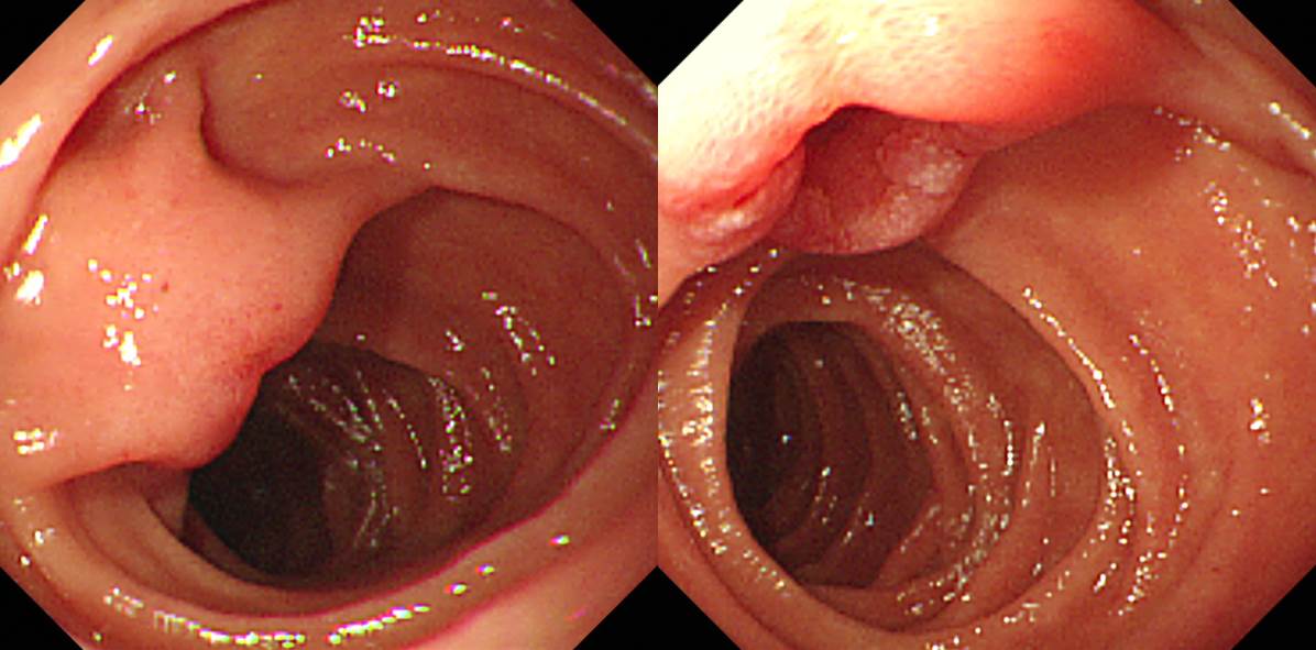
![]() 4. Ampulla of Vater cancer
4. Ampulla of Vater cancer
![]() [References]
[References]
1) SMC Endoscopy Unit ЛяМКМПяКДПј ГЛНУАцНЧ
2) SMC Monday GI conference ЛяМКМПяКДПј РЯПјГЛНУАцБГНЧ ПљПфСЁНЩМвШБтС§ДуШИ
3) SMC Thursday endoscopy conference ЛяМКМПяКДПј РЯПјГЛНУАцБГНЧ ИёПфСЁНЩГЛНУАцС§ДуШИ
© РЯПјГЛНУАцБГНЧ ЙйИЅГЛНУАцПЌБИМв РЬСиЧр. EndoTODAY Endoscopy Learning Center. Lee Jun Haeng.